. 2000 Nov 21;97(24):13057-62.
doi: 10.1073/pnas.97.24.13057.

Proteasome inhibition interferes with gag polyprotein processing, release, and maturation of HIV-1 and HIV-2

Affiliations

Proteasome inhibition interferes with gag polyprotein processing, release, and maturation of HIV-1 and HIV-2

U Schubert et al. Proc Natl Acad Sci U S A. .

Abstract

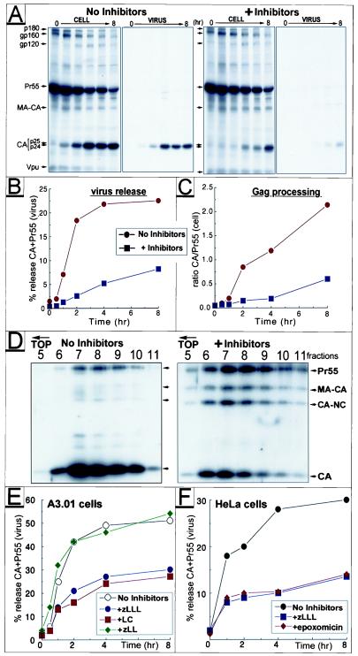
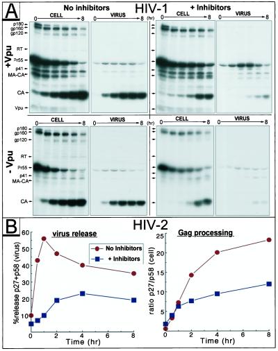
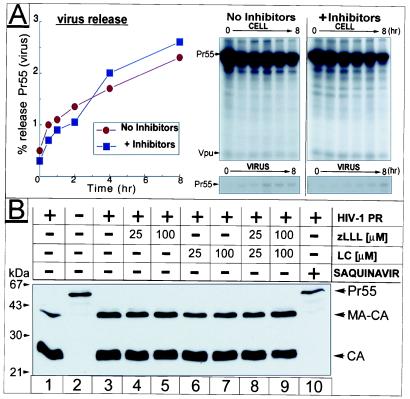
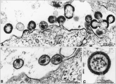
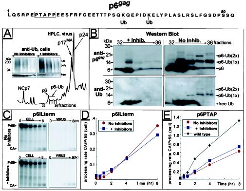
Retrovirus assembly and maturation involve folding and transport of viral proteins to the virus assembly site followed by subsequent proteolytic cleavage of the Gag polyprotein within the nascent virion. We report that inhibiting proteasomes severely decreases the budding, maturation, and infectivity of HIV. Although processing of the Env glycoproteins is not changed, proteasome inhibitors inhibit processing of Gag polyprotein by the viral protease without affecting the activity of the HIV-1 viral protease itself, as demonstrated by in vitro processing of HIV-1 Gag polyprotein Pr55. Furthermore, this effect occurs independently of the virus release function of the HIV-1 accessory protein Vpu and is not limited to HIV-1, as proteasome inhibitors also reduce virus release and Gag processing of HIV-2. Electron microscopy analysis revealed ultrastructural changes in budding virions similar to mutants in the late assembly domain of p6(gag), a C-terminal domain of Pr55 required for efficient virus maturation and release. Proteasome inhibition reduced the level of free ubiquitin in HIV-1-infected cells and prevented monoubiquitination of p6(gag). Consistent with this, viruses with mutations in PR or p6(gag) were resistant to detrimental effects mediated by proteasome inhibitors. These results indicate the requirement for an active proteasome/ubiquitin system in release and maturation of infectious HIV particles and provide a potential pharmaceutical strategy for interfering with retrovirus replication.


Figures

Figure 1
Figure 1
Effect of proteasome inhibitors on HIV-1 release and Gag processing. HeLa cells transfected with HIV-1NL4–3 were treated with 10 μM each of zLLL and LC or were left untreated during a pulse–chase experiment. Viral proteins were immunoprecipitated from the cell lysates, pelleted virions, and clarified supernatant, separated by SDS/PAGE, and analyzed by fluorography (A). Positions of the two major CA products, p24 and p25, are indicated by double arrows. The time course of virus release was calculated as the percentage of Gag (Pr55 and CA) present in the virus pellet relative to the total amount of Gag detected intra- and extracellularly (B). The rate of Pr55 processing was estimated by calculating the ratio of CA vs. Pr55 detected intracellularly at different time points (C). D shows a sucrose density gradient analysis of virus particles produced from HIV-1-infected A3.01 cells in the presence or absence of zLLL/LC. Individual fractions of the gradient were analyzed by Western blot by using HIV-1-specific antiserum. For studies on proteasome specificity, infected A3.01 (E) or transfected HeLa cells (F) were treated individually with proteasome inhibitors zLLL, LC, and epoxomicine or the control inhibitor zLL, respectively (final concentration 10 μM). After pulse–chase, virus release was calculated as above.
Figure 2
Figure 2
Proteasome inhibitors interfere with Gag processing and release of HIV-1 and HIV-2 in a Vpu-independent manner. A3.01 cells infected with HIV-1NL4–3 (+Vpu) or the Vpu-mutant vpuDEL-1 (−Vpu) (A), and HeLa cells transfected with the HIV-2 proviral plasmid pROD10 (B) were incubated in the presence or absence of inhibitors (10 μM each of zLLL and LC) and subjected to pulse–chase studies. In B, the HIV-2 Gag precursor Pr58 and the major processing product p27CA collected by immunoprecipitation were quantitated, and the time course of particle release and efficiency of intracellular Gag processing were calculated as described for Fig. 1.
Figure 3
Figure 3
Effect of proteasome inhibitors on PR activity. In A, a pulse–chase similar to Fig. 1 was conducted in HeLa cells transfected with the PR mutant pD25A. Relevant parts of the fluorograms depicting cell and virus fractions are shown (Right), and calculation of release kinetics is depicted (Left). (B) Recombinant Pr55 and PR were incubated in the presence or absence of inhibitors at the concentrations indicated. Cleavage reactions were analyzed by Western blot by using HIV-1 CA-specific antiserum.
Figure 4
Figure 4
Electron microscopy analysis of proteasome inhibitor-treated HIV-1-infected cells. HIV-1-infected MT-4 cells were treated with 50 μM zLLL for 5 h and fixed for thin-section electron microscopy. A shows an overview of infected cells with budding structures and immature virus. B shows mature extracellular HIV-1 particles, and C shows a higher-magnification view of an immature particle still connected to the cellular membrane.
Figure 5
Figure 5
Variants of HIV-1 with alterations in p6gag are insensitive to proteasome inhibitors that prevent monoubiquitination in wild-type p6gag. Proteins from purified HIV-1NL4–3 particles produced in H9 cells in the presence or absence of 25 μM zLLL/LC were separated by HPLC. The HPLC A206 profile is shown in A. Increasing aliquots, 5–30 μl of lysates of cells used for virus production, were analyzed by Western blot by using monoclonal antibody detecting both poly- and mono-Ub (A Inset). Selected HPLC fractions of separated virions were analyzed by Western blot (B) by using antibodies specific for Ub, followed by stripping and reprobing with anti-p6gag. Positions of free Ub and p6gag as well as those of mono- [p6-Ub (1×)] and di- [p6-Ub (2×)] ubiquitinated conjugates are indicated (Right). HeLa cells transfected with p6ILterm (C and D) or p6PTAP (E) were subjected to treatment with 25 μM zLLL/LC, pulse–chase experiments were conducted as described for Figs. 1–3, and rates of Gag processing were calculated (D and E). Only relevant parts of fluorograms depicting cell and virus fractions for p6ILterm-transfected cells are shown in C. In E, rate of Gag processing established for wild-type HIV-1NL4–3 in cells not treated with inhibitors was included for comparison. Amino acid sequence of the p6gag domain derived from HIV-1NL4–3 is indicated (Top).

Comment in

References

    1. Hershko A, Ciechanover A. Annu Rev Biochem. 1998;67:425–479. - PubMed
    1. Shih S C, Sloper-Mould K E, Hicke L. EMBO J. 2000;19:187–198. - PMC - PubMed
    1. Schubert U, Antón L C, Bacik I, Cox J H, Bour S, Bennink J R, Orlowski M, Strebel K, Yewdell J W. J Virol. 1998;72:2280–2288. - PMC - PubMed
    1. Ott D E, Coren L V, Copeland T D, Kane B P, Johnson D G, Sowder C, 2nd, Yoshinaka Y, Oroszlan S, Arthur L O, Henderson L E. J Virol. 1998;72:2962–2968. - PMC - PubMed
    1. Putterman D, Pepinsky R B, Vogt V M. Virology. 1990;176:633–637. - PubMed

Publication types

MeSH terms

Substances