. 2000 Nov 21;97(24):13063-8.
doi: 10.1073/pnas.97.24.13063.

A role for ubiquitin ligase recruitment in retrovirus release

Affiliations

A role for ubiquitin ligase recruitment in retrovirus release

B Strack et al. Proc Natl Acad Sci U S A. .

Abstract

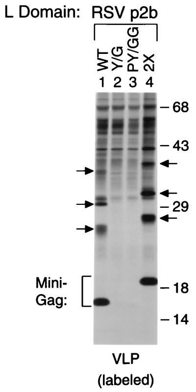
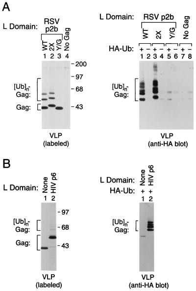
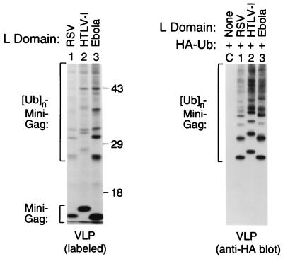
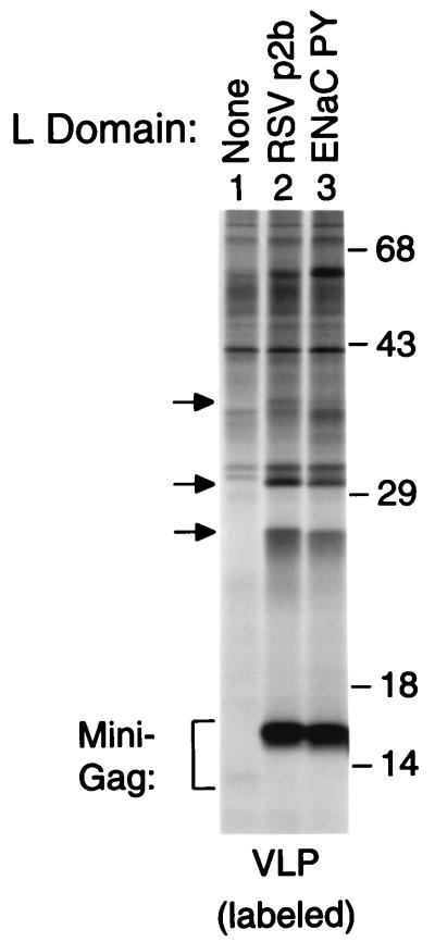
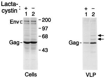
Retroviral Gag polyproteins have specific regions, commonly referred to as late assembly (L) domains, which are required for the efficient separation of assembled virions from the host cell. The L domain of HIV-1 is in the C-terminal p6(gag) domain and contains an essential P(T/S)AP core motif that is widely conserved among lentiviruses. In contrast, the L domains of oncoretroviruses such as Rous sarcoma virus (RSV) have a more N-terminal location and a PPxY core motif. In the present study, we used chimeric Gag constructs to probe for L domain activity, and observed that the unrelated L domains of RSV and HIV-1 both induced the appearance of Gag-ubiquitin conjugates in virus-like particles (VLP). Furthermore, a single-amino acid substitution that abolished the activity of the RSV L domain in VLP release also abrogated its ability to induce Gag ubiquitination. Particularly robust Gag ubiquitination and enhancement of VLP release were observed in the presence of the candidate L domain of Ebola virus, which contains overlapping P(T/S)AP and PPxY motifs. The release defect of a minimal Gag construct could also be corrected through the attachment of a peptide that serves as a physiological docking site for the ubiquitin ligase Nedd4. Furthermore, VLP formation by a full-length Gag polyprotein was sensitive to lactacystin, which depletes the levels of free ubiquitin through inhibition of the proteasome. Our findings suggest that the engagement of the ubiquitin conjugation machinery by L domains plays a crucial role in the release of a diverse group of enveloped viruses.


Figures

Figure 1
Figure 1
An intact RSV L domain rescues VLP formation by a minimal Gag construct and induces the incorporation of modified Gag molecules. HeLa cells were transfected with Δ-ZWT-p2b, which encodes a chimeric minimal Gag molecule with a WT copy of RSV p2bgag at the C terminus (Lane 1), or with the indicated variants of Δ-ZWT-p2b (Lanes 2–4). The Y/G variant (Lane 2) has the single tyrosine in p2bgag changed to glycine, the PY/GG variant (Lane 3) has both the tyrosine and the preceding proline replaced by glycine residues, and the 2× variant (Lane 4) has two tandem copies of p2bgag at the C terminus. After metabolic labeling of the transfected cells with [35S]methionine, VLPs released into the medium were pelleted through sucrose and analyzed by SDS/PAGE. The arrows indicate the positions of protein species that migrated more slowly than the expected minimal Gag molecules. The positions of migration of molecular mass markers (in kilodaltons) are indicated on the right.
Figure 2
Figure 2
Induction of Gag ubiquitination by functional L domains. 293T (A) or HeLa (B) cells were transfected with proviral constructs expressing variants of the L domain-independent ZWT Gag molecule that have either WT p2bgag, 2× p2bgag, or Y/G p2bgag (A) or p6gag (B) attached to the C terminus. As controls, a proviral construct unable to express Gag (A) and the unmodified ZWT construct (B) were used. To compare the levels of VLP formation, particulate material released into the medium during metabolic labeling with [35S]methionine was pelleted through sucrose and analyzed directly by SDS/PAGE (Left panels). To detect ubiquitin conjugates, each Gag construct was cotransfected with an expression vector for HA-tagged ubiquitin (HA-Ub), or with the empty vector, and sucrose-purified VLPs were analyzed by immunoblotting with a HA-specific monoclonal antibody (Right panels).
Figure 3
Figure 3
Potent L domain activity and ubiquitin ligase recruitment by HTLV-I and Ebola virus peptides with combined PPxY and P(T/S)AP motifs. HeLa cells were transfected with proviral constructs expressing the L domain-dependent minimal Gag molecule Δ-ZWT (Lane C) or variants of Δ-ZWT that contain at the C terminus either RSV p2bgag or the candidate L domains of HTLV-I or Ebola virus listed in Table 1 (Lanes 1–3). Unless an L domain is added, the Δ-ZWT molecule forms negligible amounts of VLP (see Fig. 4). VLPs released during metabolic labeling were pelleted through sucrose and analyzed directly by SDS/PAGE (Left). To confirm the presence of ubiquitin conjugates in VLP, the Gag constructs were cotransfected with an expression vector for HA-tagged ubiquitin, and sucrose-purified VLPs were analyzed by immunoblotting with a HA-specific monoclonal antibody (Right).
Figure 4
Figure 4
A physiological interaction site for the ubiquitin ligase Nedd4 can act as an L domain. HeLa cells were transfected with proviral constructs expressing the L domain-dependent minimal Gag molecule Δ-ZWT (lane 1), or variants of Δ-ZWT that contain at the C terminus either RSV p2bgag (lane 2) or the sequence TAPPPAYATLG from the α subunit of the human ENaC (lane 3). To compare the levels of VLP formation, particulate material released into the medium during metabolic labeling with [35S]methionine was pelleted through sucrose and analyzed directly by SDS/PAGE. In lane 1, the faint band just below the position of the 14 kDa molecular mass marker corresponds to the parental Δ-ZWT molecule. The arrows indicate the positions of Gag-ubiquitin conjugates.
Figure 5
Figure 5
Inhibition of VLP formation by lactacystin. HeLa cells were transfected with the chimeric proviral construct HXBH10/SIVgag, which expresses the full-length Gag polyprotein of SIVmac239 in the absence of a viral protease. The cells were split 48 h posttransfection and kept for 2 h in the presence or absence of 10 μM lactacystin, followed by 4 h of radiolabeling with [35S]methionine in the absence of lactacystin. The cells were then lysed, and cell-associated viral proteins were immunoprecipitated and resolved by SDS/PAGE (Left). VLPs released during the 4-h labeling period were pelleted through sucrose and analyzed directly by SDS/PAGE (Right). The arrows indicate the expected positions of mono- and di-ubiquitinated forms of Gag.

Comment in

References

    1. Gottlinger H G, Dorfman T, Sodroski J G, Haseltine W A. Proc Natl Acad Sci USA. 1991;88:3195–3199. - PMC - PubMed
    1. Huang M, Orenstein J M, Martin M A, Freed E O. J Virol. 1995;69:6810–6818. - PMC - PubMed
    1. Puffer B A, Parent L J, Wills J W, Montelaro R C. J Virol. 1997;71:6541–6546. - PMC - PubMed
    1. Wills J W, Cameron C E, Wilson C B, Xiang Y, Bennett R P, Leis J. J Virol. 1994;68:6605–6618. - PMC - PubMed
    1. Xiang Y, Cameron C E, Wills J W, Leis J. J Virol. 1996;70:5695–5700. - PMC - PubMed

Publication types