. 2000 Nov 21;97(24):13069-74.
doi: 10.1073/pnas.97.24.13069.

Ubiquitin is part of the retrovirus budding machinery

Affiliations

Ubiquitin is part of the retrovirus budding machinery

A Patnaik et al. Proc Natl Acad Sci U S A. .

Abstract

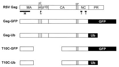
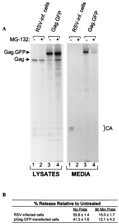
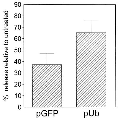
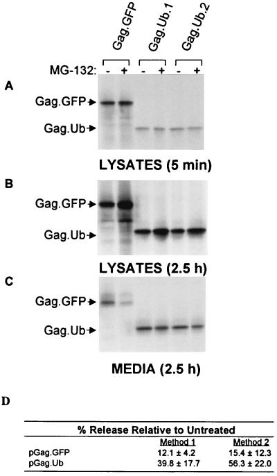
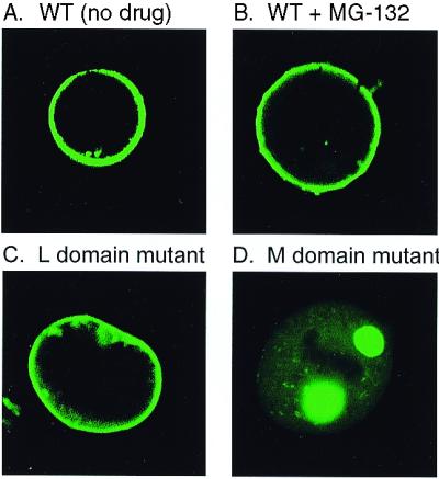
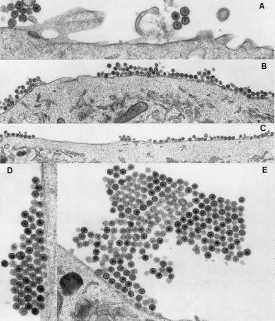
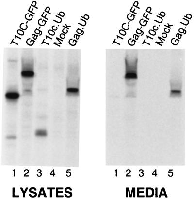
Retroviruses contain relatively large amounts of ubiquitin, but the significance of this finding has been unknown. Here, we show that drugs that are known to reduce the level of free ubiquitin in the cell dramatically reduced the release of Rous sarcoma virus, an avian retrovirus. This effect was suppressed by overexpressing ubiquitin and also by directly fusing ubiquitin to the C terminus of Gag, the viral protein that directs budding and particle release. The block to budding was found to be at the plasma membrane, and electron microscopy revealed that the reduced level of ubiquitin results in a failure of mature virus particles to separate from each other and from the plasma membrane during budding. These data indicate that ubiquitin is actually part of the budding machinery.


Figures

Figure 1
Figure 1
RSV Gag derivatives used in this study. The wild-type polyprotein is shown at the top and its proteolytic cleavage products are indicated. The domains required for budding are indicated below Gag. The M domain mediates the binding of Gag to the cytoplasmic face of the plasma membrane. The I domains provide the major regions of interaction among the 1,500 molecules that create a virion particle. The L domain is required for the virus–cell separation steps that occur late in the budding pathway. The foreign sequences in Gag-GFP, Gag-Ub, T10C-GFP, and T10C-Ub replace the protease (PR) sequence and the last six residues of the nucleocapsid (NC) sequence.
Figure 2
Figure 2
Proteasome inhibitors block RSV budding. (A) RSV-infected QT6 (quail) cells were untreated (lanes 1) or pretreated with 80 μM MG-132 for 90 min before metabolically labeling with [35S]methionine for 2.5 h, also in the presence of the drug (lanes 2). Alternatively, QT6 cells were transfected with pGag-GFP, which expresses a recombinant RSV Gag protein in which C-terminal sequences have been replaced with GFP. The next day, the transfected cells were untreated (lanes 3) or pretreated with 80 μM MG-132 (lanes 4) before labeling as above. The cells and particles in the growth medium were detergent lysed, and the Gag proteins were immunoprecipitated and electrophoresed in SDS/12% polyacrylamide gels before detection by autoradiography. (B) The negative impact of MG132 on budding was quantitated by using a PhosphorImager. Cells were either pretreated with MG-132 for 90 min in addition to treatment during the 2.5-h labeling period, or treated only during the labeling period. The budding efficiency in each culture was calculated as the amount of Gag protein in the medium (CA products) divided by the total in the lysates (Gag precursor and CA products) and media. The effects of MG-132 on budding (% release relative to untreated) were then determined by computing the ratios of budding efficiency in the absence and presence of drug. The averages from two no-pretreatment and three pretreatment experiments are shown .
Figure 3
Figure 3
Overexpression of Ub suppresses the effects of proteasome inhibitors. QT6 cells were cotransfected with 5 μg of (pGag.GFP) and 5 μg of either a human Ub expression plasmid (pUb) or the same plasmid containing the GFP-coding sequence instead (pGFP; negative control). The next day, cells were pretreated with 20 μM MG-132 for 90 min before labeling for 2.5 h with [35S]methionine, also in the presence of 20 μM MG-132. The labeled Gag proteins were collected and analyzed as described in Fig. 2. The graph shows the average of two experiments.
Figure 4
Figure 4
A Gag-Ub chimera is comparatively insensitive to proteasome inhibitors. QT6 cells were transfected with either pGag-GFP or pGag-Ub (clones 1 and 2). The next day, the cells were pretreated with 80 μM MG-132 for 90 min before labeling with [35S]methionine, also in the presence of drug. (A) Gag proteins from cells that had been labeled for only 5 min were collected to compare the initial rates of protein synthesis of the treated (95 min, total) and untreated cells. (B and C) Identical sets of plates were labeled for 2.5 h to allow budding to proceed, and Gag proteins were collected from the cell lysates and media of the treated (4 h, total) and untreated cultures, as in Fig. 1. (D) PhosphorImager analysis of the budding efficiencies of Gag-GFP and Gag-Ub in the presence of MG-132. Data from three different experiments were used to calculate the averages by two different methods. Method 1 used only the data from the plates labeled for 2.5 h, which was used to calculate the percent of the total Gag protein found in the medium in treated vs. untreated cells. Method 2 used the data from the plates labeled for only 5 min to normalize the signals in the media after 2.5 h of labeling. Both methods show that Gag-Ub is much less sensitive to the effects of MG-132.
Figure 5
Figure 5
Gag accumulates on the plasma membrane in the presence of MG-132. QT6 cells were transfected with the indicated Gag-GFP constructs, and, 24 h, later the cells were examined by confocal microscopy. (A, C, and D) Untreated cells. (B) Cells treated with 80 μM MG-132 for 3 h.
Figure 6
Figure 6
Electron microscopy of MG-132-treated RSV-infected cells. (A) DMSO-treated RSV-infected cells, magnified ×27,500. (B—E) 80 μM MG-132-treated RSV-infected cells, magnified ×13,280 (B), ×10,950 (C), ×23,300 (D), and ×12,500 (E).
Figure 7
Figure 7
Corequirement for the L domain and Ub. QT6 cells were transfected with the indicated Gag-GFP and Gag-Ub constructs, and, 24 h later, the cells were labeled for 2.5 h with [35S]methionine. No proteasome inhibitors were used in this experiment.

Comment in

References

    1. Ciechanover A, Orian A, Schwartz A L. BioEssays. 2000;22:442–451. - PubMed
    1. Hicke L. FASEB J. 1997;11:1215–1226. - PubMed
    1. Shih S C, Sloper-Mould K E, Hicke L. EMBO J. 2000;19:187–198. - PMC - PubMed
    1. Terrel J, Shih S, Dunn R, Hicke L. Mol Cell. 1998;1:193–202. - PubMed
    1. Nakatsu F, Sakuma M, Matsuo Y, Arase H, Yamasaki H, Nakamura N, Saito T, Ohno H. J Biol Chem. 2000;275:26213–26219. - PubMed

Publication types

MeSH terms

LinkOut - more resources