. 2000 Nov 21;97(24):13306-11.
doi: 10.1073/pnas.97.24.13306.

Acquired, nonrandom chromosomal abnormalities associated with the development of acute promyelocytic leukemia in transgenic mice

Affiliations

Acquired, nonrandom chromosomal abnormalities associated with the development of acute promyelocytic leukemia in transgenic mice

D B Zimonjic et al. Proc Natl Acad Sci U S A. .

Abstract

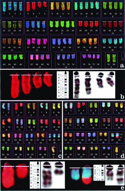
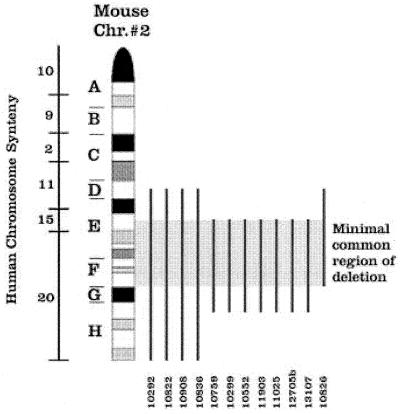
We previously generated a transgenic mouse model for acute promyelocytic leukemia (APL) by expressing the promyelocytic leukemia (PML)-retinoic acid receptor (RARalpha) cDNA in early myeloid cells. This fusion protein causes a myeloproliferative disease in 100% of animals, but only 15-20% of the animals develop acute leukemia after a long latency period (6-13 months). PML-RARalpha is therefore necessary, but not sufficient, for APL development. The coexpression of a reciprocal form of the fusion, RARalpha-PML, increased the likelihood of APL development (55-60%), but did not shorten latency. Together, these results suggested that additional genetic events are required for the development of APL. We therefore evaluated the splenic tumor cells from 18 transgenic mice with APL for evidence of secondary genetic events, by using spectral karyotyping analysis. Interstitial or terminal deletions of the distal region of one copy of chromosome 2 [del(2)] were found in 1/5 tumors expressing PML-RARalpha, but in 11/13 tumors expressing both PML-RARalpha and RARalpha-PML (P < 0.05). Leukemic cells that contained a deletion on chromosome 2 often contained additional chromosomal gains (especially of 15), chromosomal losses (especially of 11 or X/Y), or were tetraploid (P </= 0.001). These changes did not commonly occur in nontransgenic littermates, nor in aged transgenic mice that did not develop APL. These results suggest that expression of RARalpha-PML increases the likelihood of chromosome 2 deletions in APL cells. Deletion 2 appears to predispose APL cells to further chromosomal instability, which may lead to the acquisition of additional changes that provide an advantage to the transformed cells.


Figures

Figure 1
Figure 1
(a) Spectral karyotype of a tetraploid cell from mouse 10759, containing an interstitial deletion of chromosome 2 as the only structural abnormality. (b) Localization of the chromosome 2 interstitial deletion from a. The comparison of chromosome's inverted 4′,6-diamidino-2-phenylindole G-banding pattern and the G-banding ideogram for mouse chromosome 2 allows the identification of the deletion as del(2)(E2H1). (c) Spectral karyotype of a pseudodiploid tumor cell from mouse 11908, containing a deletion of chromosome 2 del(2)(Dter), an extra chromosome 15, and loss of the Y chromosome. (d) Spectral karyotype of a hypodiploid cell from mouse 10826, containing a translocation 2;15 as the only structural alteration, and loss of a sex chromosome. (e) Localization of the breakpoints of the deleted chromosome 2 and derivative 15 from d. Chromosome 2 (Left) is deleted at band 2D (as in c), and chromosome 15 (Right) at band 15E. The translocated fragment from chromosome 2 appears to span bands 2G through the terminus. Therefore, the abnormality is defined as translocation t(2;15) (G-ter;E) and most likely involves loss of chromosome 2 material containing bands 2E and 2F.
Figure 2
Figure 2
Staging of APL tumors. (A--D) Flow cytometric analysis of spleen cells from a wild-type mouse (A) or spleen cells from mice dying with APL (BD) for expression of Gr-1 and CD34. (EH) Wright-Giemsa stains of the same splenic cells shown in AD. Samples are as follows: (A and E) wild-type spleen; (B and F) a highly differentiated stage 1 tumor from mouse 10836; (C and G) an intermediate stage 2 APL tumor from mouse 13080; (D and H) an immature stage 3 APL tumor from mouse 11309.
Figure 3
Figure 3
Summary of chromosome 2 deletions. Regions of synteny between mouse chromosome 2 and human chromosomes are shown. The deleted portion of chromosome 2 is designated by the vertical line for each APL tumor. In the tumor derived from mouse 10826, the distal part of chromosome 2 (G-ter) is translocated to chromosome 15. The minimal common region of deletion for all 12 tumors therefore is confined within the region 2E2-F3.

References

    1. Lo Coco F, Diverio D, Falini B, Biondi A, Nervi C, Pelicci P G. Blood. 1999;94:12–22. - PubMed
    1. Melnick A, Licht J D. Blood. 1999;93:3167–3215. - PubMed
    1. Grisolano J L, Wesselschmidt R L, Pelicci P G, Ley T J. Blood. 1997;89:376–387. - PubMed
    1. Brown D, Kogan S, Lagasse E, Weissman I, Alcalay M, Pelicci P G, Atwater S, Bishop J M. Proc Natl Acad Sci USA. 1997;94:2551–2556. - PMC - PubMed
    1. He L-Z, Tribioli C, Rivi R, Peruzzi D, Pelicci P G, Soares V, Cattoretti G, Pandolfi P P. Proc Natl Acad Sci USA. 1997;94:5302–5307. - PMC - PubMed

Publication types

Substances

LinkOut - more resources