. 2000 Nov 21;97(24):13336-41.
doi: 10.1073/pnas.97.24.13336.

High-resolution structure of hair-cell tip links

Affiliations

High-resolution structure of hair-cell tip links

B Kachar et al. Proc Natl Acad Sci U S A. .

Erratum in

  • Proc Natl Acad Sci U S A. 2013 Jul 16;110(29):12155

Abstract

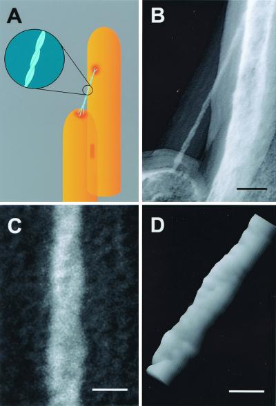
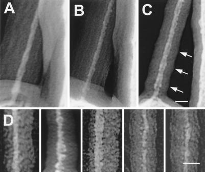
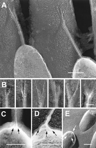
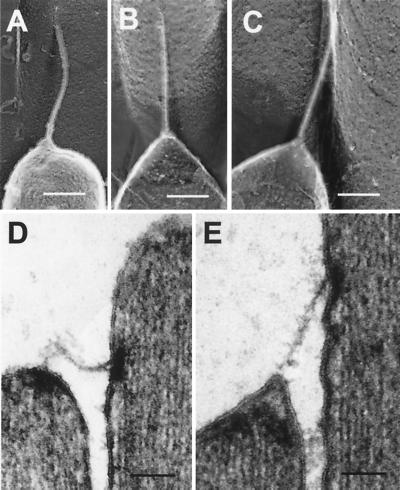
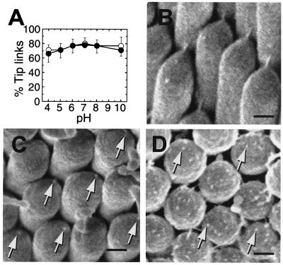
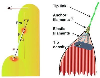
Transduction-channel gating by hair cells apparently requires a gating spring, an elastic element that transmits force to the channels. To determine whether the gating spring is the tip link, a filament interconnecting two stereocilia along the axis of mechanical sensitivity, we examined the tip link's structure at high resolution by using rapid-freeze, deep-etch electron microscopy. We found that the tip link is a right-handed, coiled double filament that usually forks into two branches before contacting a taller stereocilium; at the other end, several short filaments extend to the tip link from the shorter stereocilium. The structure of the tip link suggests that it is either a helical polymer or a braided pair of filamentous macromolecules and is thus likely to be relatively stiff and inextensible. Such behavior is incompatible with the measured elasticity of the gating spring, suggesting that the gating spring instead lies in series with the helical segment of the tip link.


Figures

Figure 1
Figure 1
Helical structure of the tip link. (A) Proposed model for tip-link structure. Two helically intertwined protofilaments (Inset) make up the tip link, attaching at two points to the taller stereocilium and contacting three filaments emanating from the shorter stereocilium. (B) Freeze-etch image of tip link from guinea pig cochlea. Note the thick carbon coat forming a halo around the tip link and the stereocilia surface. (C) Higher magnification view of the tip link in B; we suggest that the reader view this figure from a shallow angle perspective (from bottom to top) to better view the helical configuration of the tip link. We confirmed right-handedness by imaging stereo pairs with the concave side of the replica up. (D) Surface plot of the pixel intensities of the digitized image of the tip link shown in B created with National Institutes of Health image. The pseudo-three-dimensional image helped visualize the helical configuration and the possible periodic substructure of the protofilaments. (Scale bars: B = 50 nm; C and D = 10 nm.)
Figure 2
Figure 2
Variability in appearance of tip-link extended structure. (A–C) Images of the tip link of Fig. 1B acquired with different tilt angles showing different apparent thickness of the tip link and its carbon coat. Despite degradation of the platinum image of the tip link, the enveloping carbon coat still shows the 30-nm periodic widening (arrows) and narrowing consistent with the projected view of a helical structure. (D) Images of the extended structure of different tip links from guinea pig cochlea (first two panels from left to right) and from bullfrog sacculus (three panels to the right). (Scale bars = 15 nm.)
Figure 3
Figure 3
Upper and lower attachments of the tip link. (A and B) Freeze-etch images of tip-link upper insertions in guinea pig cochlea (A) and (left to right) two from guinea pig cochlea, two from bullfrog sacculus, and two from guinea pig utriculus (B). Each example shows pronounced branching. (C and D) Freeze-etch images of the tip-link lower insertion in stereocilia from bullfrog sacculus (C) and guinea pig utriculus (D); multiple strands (arrows) arise from the stereociliary tip. (E) Freeze-fracture image of stereociliary tips from bullfrog sacculus; indentations at tips are indicated by arrows. (Scale bars: A = 100 nm, B = 25 nm; C–E = 100 nm.)
Figure 4
Figure 4
Response of the tip link to compressive and tensile forces. (A) Freeze-etch images of a smoothly buckled bullfrog tip link. (B and C) Freeze-etch images of a bullfrog tip link apparently under tension; note that lower membrane is cone-shaped, suggesting that tenting is present. (D) Thin-section TEM image of a tip link in a relaxed state. (E) Thin-section TEM image of a tip link in a tensed state. The membrane appears to pull away from the tip density. (Scale bars = 100 nm.)
Figure 5
Figure 5
Response of tip links to chemical treatment. (A) Chicken basilar papillae were exposed to solutions of varying pH and either 4 mM (●) or 1.4 μM (○) CaCl2. For each point, tip links from 14–16 hair bundles from 3–4 papillae were counted from SEM images; each point corresponds to ≈1,000 tip-link positions. Means ± standard error are plotted. Bundles were chosen from all regions of the papilla. (B–D) Field-emission SEM images of chicken basilar-papilla hair bundles before and after BAPTA treatment. (B) Control. (C) Immediately after BAPTA treatment; remnants are seen where tip links formerly anchored (arrows). (D) 2 h after BAPTA treatment, during tip-link regeneration. Protuberances on the surface of each stereociliary tip are indicated by arrows. [Scale bars: B = 200 nm (applies to C and D).]
Figure 6
Figure 6
Models for tip-link structure. (Left) Model indicating buckling forces during tip-link compression. At the buckling force F, the force pushing the upper insertion point parallel to the membrane is Fm = F cos(α), where α is the angle formed by the tip link and the stereocilium (≈30°). (Right) Model for tip-link structure demonstrating responses to tip-link extension; note tenting of membrane and extended putative elastic filaments located between the membrane and the osmiophilic tip density.

References

    1. Hudspeth A J. Nature (London) 1989;341:397–404. - PubMed
    1. Hudspeth A J. Soc Gen Physiol Ser. 1992;47:357–370. - PubMed
    1. Pickles J O, Comis S D, Osborne M P. Hearing Res. 1984;15:103–112. - PubMed
    1. Assad J A, Shepherd G M G, Corey D P. Neuron. 1991;7:985–994. - PubMed
    1. Zhao Y, Yamoah E N, Gillespie P G. Proc Natl Acad Sci USA. 1996;93:15469–15474. - PMC - PubMed

Publication types

LinkOut - more resources