. 2000 Nov 21;97(24):13360-5.
doi: 10.1073/pnas.97.24.13360.

Remodeling of alpha-amino-3-hydroxy-5-methyl-4-isoxazole-propionic acid receptor subunit composition in hippocampal neurons after global ischemia

Affiliations

Remodeling of alpha-amino-3-hydroxy-5-methyl-4-isoxazole-propionic acid receptor subunit composition in hippocampal neurons after global ischemia

T Opitz et al. Proc Natl Acad Sci U S A. .

Erratum in

  • Proc Natl Acad Sci U S A 2001 Jan 30;98(3):1317. Optiz T [corrected to Opitz T]

Abstract

Transient global ischemia induces selective delayed cell death, primarily of principal neurons in the hippocampal CA1. However, the molecular mechanisms underlying ischemia-induced cell death are as yet unclear. The present study shows that global ischemia triggers a pronounced and cell-specific reduction in GluR2 [the subunit that limits Ca(2+) permeability of alpha-amino-3-hydroxy-5-methyl-4-isoxazole-propionic acid (AMPA) receptors] in vulnerable CA1 neurons, as evidenced by immunofluorescence of brain sections and Western blot analysis of microdissected hippocampal subfields. At 72 h after ischemia (a time before cell death), virtually all CA1 pyramidal neurons exhibited greatly reduced GluR2 immunolabeling throughout their somata and dendritic processes. GluR2 immunolabeling was unchanged in pyramidal cells of the CA3 and granule cells of the dentate gyrus, regions resistant to ischemia-induced damage. Immunolabeling of the AMPA receptor subunit GluR1 was unchanged in CA1, CA3, and dentate gyrus. Western analysis indicated that GluR2 subunit abundance was markedly reduced in CA1 at 60 and 72 h after the ischemic insult; GluR1 abundance was unchanged in all subfields at all times examined. These findings, together with the previous observation of enhanced AMPA-elicited Ca(2+) influx in postischemic CA1 neurons, show that functional GluR2-lacking, Ca(2+)-permeable AMPA receptors are expressed in vulnerable neurons before cell death. Thus, the present study provides an important link in the postulated causal chain between global ischemia and delayed death of CA1 pyramidal neurons.


Figures

Figure 1
Figure 1
Global ischemia induces selective, delayed neurodegeneration in hippocampal CA1. Toluidine blue staining of plastic-embedded sections through the dorsal hippocampus. (A, D, and G) Control brain from an animal killed at 7 days after sham operation. (B, E, and H) At 72 h after transient (5 min) global ischemia there was no histologically detectable neuronal death in any hippocampal subfield. (C, F, and I) By 7 days after ischemia, the pyramidal cell layer of CA1 exhibited essentially complete loss of neurons, whereas CA3 and DG showed no damage.
Figure 2
Figure 2
Global ischemia induces cell-specific suppression of GluR2, but not GluR1, immunolabeling in CA1 pyramidal neurons. GluR2 (Upper) and GluR1 (Lower) immunolabeling in the CA1 pyramidal cell layer (A–D), the CA3a pyramidal cell layer (E–H) and DG granule cell layer (I–L) in sections from a control animal (A, C, E, G, I, and K) and an experimental animal 72 h after ischemia (B, D, F, H, J, and L). Data are typical of two control and four ischemic animals. Ischemia induced down-regulation of GluR2, but not GluR1, in CA1 pyramidal neurons. Ischemia did not alter GluR1 or GluR2 immunolabeling in CA3a pyramidal neurons or DG granule cells. so: stratum oriens; sp: stratum pyramidale; sr: stratum radiatum; slu: stratum lucidum; ml: molecular layer; gcl: granule cell layer; h: hilus.
Figure 3
Figure 3
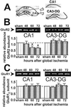
Global ischemia induces subunit-specific down-regulation of GluR2 abundance in CA1. (A) Microdissection of the hippocampus for Western blot analysis. (B) Representative Western blots probed with GluR2C Ab (Upper) and relative GluR2 subunit abundance (defined as the ratio of band densities for experimental vs. control samples; Lower) for CA1 (Left) and CA3-DG (Right) in control and postischemic animals. Protein samples of hippocampal CA1 and CA3-DG were from control animals at 72 h and experimental animals at 48, 60, and 72 h after brief (5 min) global ischemia. Relative GluR2 subunit abundance was markedly decreased in CA1 at 60 h (to 61.5 ± 11.9% of control; n = 6 per time point; P < 0.05) and 72 h after ischemia (to 57.6 ± 7.9% of control, P < 0.05; n = 6 per time point). GluR2 subunit abundance was unchanged in CA3-DG. (C) Western blots probed with GluR1C Ab (Upper) and relative GluR1 subunit abundance displayed as in B. Aliquots of the same protein samples (hippocampal CA1 and CA3-DG) used in B were run on different gels. GluR1 subunit abundance was not altered in the CA1 or CA3-DG at any times examined after ischemia (n = 6 per time point). Band densities were corrected for protein concentration. Bars are means ± SEMs. Asterisks indicate significant difference from control values (P < 0.05).
Figure 4
Figure 4
The GluR1 C terminus is not cleaved in CA1 after global ischemia. The ratio of GluR1 subunit abundance in CA1 (Left) and CA3-DG (Right) calculated from Westerns probed with GluR1C Ab or GluR1N Ab. Protein samples were from control animals and experimental animals at 48, 60, and 72 h after ischemia. In CA1 and CA3-DG, the GluR1c/GluR1N ratio was not significantly changed at any times examined after ischemia, indicating stability of the GluR1 C terminus after ischemia. Band densities were corrected for protein concentration and normalized to corresponding control values. Bars are means ± SEMs from four experiments.

Similar articles

Cited by

References

    1. Choi D W. Trends Neurosci. 1995;18:58–60. - PubMed
    1. Pulsinelli W A, Brierley J B, Plum F. Ann Neurol. 1982;11:491–498. - PubMed
    1. Seeburg P H. Trends Neurosci. 1993;16:359–365. - PubMed
    1. Verdoorn T A, Burnashev N, Monyer H, Seeburg P H, Sakmann B. Science. 1991;252:1715–1718. - PubMed
    1. Geiger J R, Melcher T, Koh D S, Sakmann B, Seeburg P H, Jonas P, Monyer H. Neuron. 1995;15:193–204. - PubMed

Publication types

Substances