. 2000 Dec;74(24):11447-55.
doi: 10.1128/jvi.74.24.11447-11455.2000.

In vitro reconstitution of a functional duck hepatitis B virus reverse transcriptase: posttranslational activation by Hsp90

Affiliations

In vitro reconstitution of a functional duck hepatitis B virus reverse transcriptase: posttranslational activation by Hsp90

J Hu et al. J Virol. 2000 Dec.

Abstract

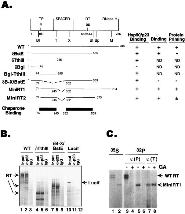
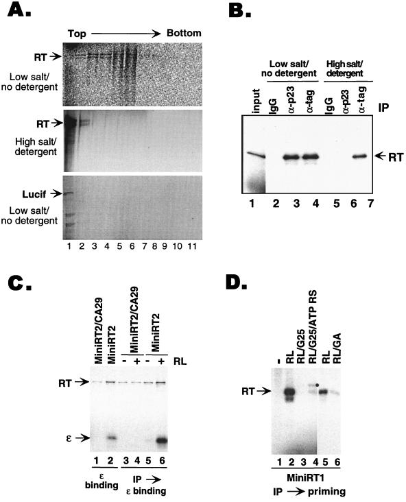
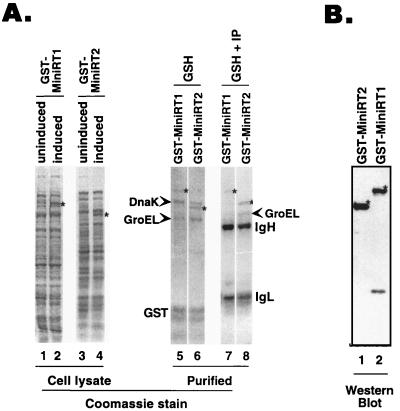
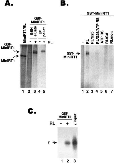
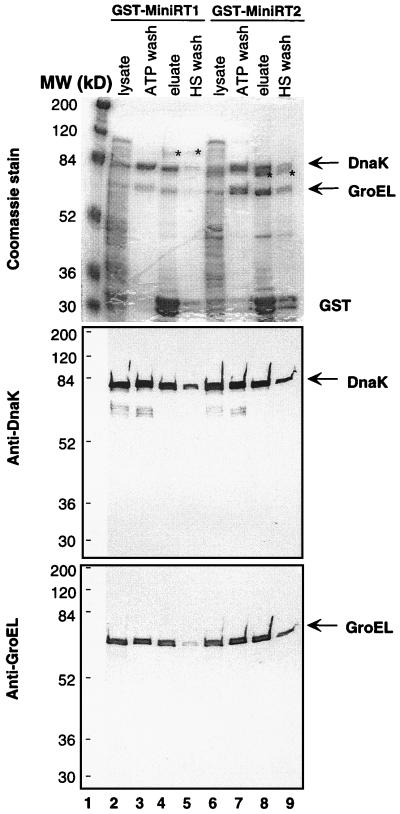
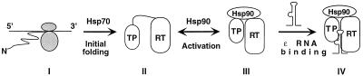
Reverse transcription in hepatitis B viruses is initiated through a unique protein priming mechanism whereby the viral reverse transcriptase (RT) first assembles into a ribonucleoprotein (RNP) complex with its RNA template and then initiates DNA synthesis de novo using the RT itself as a protein primer. RNP formation and protein priming require the assistance of host cell factors, including the molecular chaperone heat shock protein 90 (Hsp90). To better understand the mechanism of RT activation by Hsp90, we have now mapped the minimal RT sequences of the duck hepatitis B virus that are required for chaperone binding, RNP formation, and protein priming. Furthermore, we have reconstituted in vitro both RNP formation and protein priming using purified RT proteins and host factors. Our results show that (i) Hsp90 recognizes two independent domains of the RT, both of which are necessary for RNP formation and protein priming; (ii) Hsp90 function is required not only to establish, but also to maintain, the RT in a state competent for RNA binding; and (iii) Hsp90 is not required during RT synthesis and can activate the RT posttranslationally. Based on these findings, we propose a model for Hsp90 function whereby the chaperone acts as an active interdomain bridge to bring the two RT domains into a poised but labile conformation competent for RNP formation. It is anticipated that the reconstitution system established here will facilitate the isolation of additional host factors required for RT functions and further elucidation of the mechanisms of RT activation.


Figures

FIG. 1
FIG. 1
RT sequences required for chaperone association, ɛ binding, and protein priming. (A) Schematic diagram (top) of the domain structure of the DHBV RT. The primer tyrosine (residue 96) in the TP domain and the two aspartic acids (residues 513 and 514) at the RT active site are indicated, as are the unique restriction sites used to construct the various deletion mutants: Bl, BglII; Bp, BspHI; Bt, BstEII; M, MscI; T, Tth111I; X, XhoI. Below are the activities of the wild-type (WT) RT and the various truncation and deletion RT mutants in chaperone binding, ɛ binding, and protein priming, as determined by coimmunoprecipitation and in vitro protein priming assays (see Materials and Methods). The two independent chaperone-binding regions of the RT are shown schematically. ND, not determined. (B) Examples of coimmunoprecipitation of full-length RT and deletion variants of the RT with p23. δTth111I harbored the N-terminal chaperone-binding region in the TP domain, and δB-X/BstE contained the central chaperone-binding region in the RT domain (A). The RT proteins, along with the luciferase protein (Lucif; used as a negative control for immunoprecipitation), were translated in reticulocyte lysate, and the translation reaction mixtures were subjected to immunoprecipitation with a MAB (clone JJ3) specific for p23 (α-p23; lanes 2, 5, 8, and 11) or a nonimmune control antibody (IgG; lanes 3, 6, 9, and 12). The immunoprecipitates, along with the translation reaction products (input; lanes 1, 4, 7, and 10), were resolved by SDS-PAGE and the 35S-labeled RT proteins were detected by autoradiography. (C) WT RT and MiniRT1 were translated in reticulocyte lysate, and their protein priming activities were assessed by the in vitro priming assay, with or without treatment with Hsp90 inhibitor geldanamycin (GA). Lanes 1 and 2, 35S-labeled translation product (lane 1, WT RT; lane 2, MiniRT1); lanes 3 to 8, 32P-labeled WT RT (lanes 3 and 4) and mini-RT (lanes 5 to 8) as a result of the protein priming reaction in the presence of [α-32P]dATP. Following translation but before the priming reaction, aliquots of the translation reaction mixture, as indicated, were treated with GA (100 μg/ml). ɛ was added during either the translation [i.e., before GA treatment; ɛ(T)] or priming [i.e., after GA treatment; ɛ(P)] step.
FIG. 2
FIG. 2
Purification of the RT following in vitro translation and reconstitution of ɛ binding and protein priming. (A) Dissociation of the RT complexes. 35S-labeled RT (upper and middle sections) and luciferase (Lucif) (lower section) were synthesized in reticulocyte lysate and centrifuged over a linear 20 to 40% sucrose gradient at 40,000 rpm for 4 h in an SW41 rotor. For the RT sample shown in the middle section, the translation reaction mixture was first treated with 1 M NaCl and 1% NP-40 before centrifugation over the sucrose gradient made in the same high-salt and -detergent buffer. Individual fractions were then collected and resolved by SDS-PAGE. The direction of centrifugation is indicated. (B) Dissociation of RT-p23 binding. MiniRT1, tagged with the c-Myc and HA epitopes, was expressed in TnT reticulocyte lysate (lane 1) and immunoprecipitated (IP) with either a control IgG (lanes 2 and 5), an anti-p23 MAb (lanes 3 and 6), or a mixture of anti-c-Myc and anti-HA (lanes 4 and 7) antibodies. The binding and washing buffers contained either 50 mM phosphate buffer–10 mM sodium molybdate (lanes 2 to 4) or 0.5 M NaCl–0.2% NP-40 (lanes 5 to 7). The immunoprecipitated, 35S-labeled mini-RT was then resolved by SDS-PAGE and detected by autoradiography. (C) Immunoaffinity purification of mini-RT proteins and reconstitution of ɛ binding. MiniRT2 and the corresponding ɛ-binding-defective mutant, MiniRT2/CA29, were expressed in TnT reticulocyte lysate supplemented with [35S]methionine and purified by immunoprecipitation under high-salt and -detergent conditions (as described for panel B). 32P-labeled ɛ RNA was then incubated with the purified RT proteins with (lanes 4 and 6) or without (lanes 3 and 5) the addition of reticulocyte lysate. Unbound RNA was washed away, and the bound ɛ was detected following SDS-PAGE (lanes 3 to 6). As controls, labeled ɛ RNA was added to the translation reaction mixtures and thus allowed to bind to RT during translation; the RT-ɛ complex was then immunoprecipitated and detected by SDS-PAGE and autoradiography (lanes 1 and 2). (D) Reconstitution of protein priming. In vitro-translated MiniRT1 was isolated by immunoprecipitation under high-salt and -detergent conditions. The ɛ RNA was then added to the purified RT to initiate protein priming. The 32P-labeled priming reaction products were detected by autoradiography following SDS-PAGE. The priming reactions were carried out with the following supplement: nonsupplemented control (lane 1), unfractionated reticulocyte lysate (RL; lanes 2 and 5), reticulocyte lysate desalted by passing through a Sephadex G-25 column (RL/G25; lane 3), desalted reticulocyte lysate supplemented with an ATP regenerating system (see Methods and Materials) (RL/G25/ATP RS; lane 4), reticulocyte lysate plus geldanamycin (100 μg/ml) (RL/GA; lane 6). Solid circle (lane 4) reaction product observed sometimes under the indicated conditions, the nature of which is currently unknown.
FIG. 3
FIG. 3
Bacterial expression and purification of mini-RT. (A) Two GST mini-RT fusion proteins, GST-MiniRT1 and GST-MiniRT2, were expressed in BL21 cells as described in Materials and Methods. Uninduced (lanes 1 and 3) and induced (lanes 2 and 4) bacterial lysates were prepared. The fusion proteins were first purified using glutathione-agarose beads (lanes 5 and 6) and then further purified by immunoprecipitation (IP) with the anti-c-Myc antibody (lanes 7 and 8). The lysate and purified protein samples were resolved by SDS-PAGE and stained with Coomassie blue. Stars, full-length mini-RT fusion proteins; arrowheads, two major copurifying bacterial proteins (DnaK and GroEL; see Fig. 5). The Ig heavy (IgH) and light chains (IgL) and GST are also indicated. (B) GST-MiniRT1 (lane 2) and GST-MiniRT2 (lane 1) purified on glutathione-agarose beads were resolved by SDS-PAGE and detected by Western blot analysis using the anti-c-Myc antibody. Stars, full-length mini-RT fusion proteins.
FIG. 4
FIG. 4
Reconstitution of RNA binding and protein priming with mini-RT purified from bacteria. (A) GST-MiniRT1, purified either by glutathione-agarose beads (lanes 2 and 3) or by glutathione beads plus the second step of immunoaffinity purification (lanes 4 and 5), was used for the in vitro protein priming reaction in the presence of ɛ and [α-32P]dATP, with (lanes 3 and 5) or without (lanes 2 and 4) supplementation with reticulocyte lysate (RL). As a positive control, MiniRT1 (without the GST fusion) was translated in reticulocyte lysate and assayed in the priming reaction (lane 1). The 32P-labeled RT was then detected by SDS-PAGE and autoradiography. (B) Purified GST-MiniRT1 was used in the protein priming reaction as described for panel A, with the indicated supplements. Abbreviations are as defined in the Fig. 2D legend. RL/no ɛ, priming reaction mixture supplemented with reticulocyte lysate but without ɛ RNA. (C) Purified GST-MiniRT2 was used in the ɛ RNA binding assay as described for Fig. 2C, except that GST-MiniRT2 was bound to glutathione-agarose beads. 32P-labeled ɛ RNA precipitated by the RT-bound beads was then detected by SDS-PAGE and autoradiography. Reticulocyte lysate was added as indicated. Lane 3, RNA input.
FIG. 5
FIG. 5
Association of DnaK and GroEL with RT. GST-MiniRT1 and -MiniRT2 were expressed in BL21 cells and purified by using glutathione beads. The bacterial lysate (lysate) and different fractions from the purification protocol were then analyzed by Coomassie staining (top) or Western blotting using anti-DnaK (middle) or anti-GroEL (bottom). ATP wash, proteins washed off the beads following a 30-min incubation with ATP (5 mM) at room temperature; eluate, proteins eluted from the beads by glutathione; HS (high salt) wash, proteins washed off the beads by 3 M NaCl (used to regenerate the affinity resin). Stars, mini-RT fusion proteins; arrows, DnaK and GroEL.
FIG. 6
FIG. 6
Model of RT folding and activation by cellular chaperones. The nascent RT polypeptide (I) exiting the translating ribosomes is first recognized by Hsp70 (or DnaK when expressed in bacteria). With Hsp70 assistance, the TP and RT domains can then partially fold (initial folding; II), independent of each other. However, the RT remains incompetent for ɛ binding. The Hsp90 complex then recognizes the partially folded TP and RT domains and brings them together to establish an RT conformation able to bind ɛ (activation; III). This RT conformation is intrinsically unstable and requires the continued assistance of Hsp90 to maintain it. Finally, ɛ binding stabilizes this RT conformation while Hsp90 remains bound to the RT (IV).

Similar articles

Cited by

References

    1. Bartenschlager R, Junker-Niepmann M, Schaller H. The P gene product of hepatitis B virus is required as a structural component for genomic RNA encapsidation. J Virol. 1990;64:5324–5332. - PMC - PubMed
    1. Bartenschlager R, Kuhn C, Schaller H. Expression of the P-protein of the human hepatitis B virus in a vaccinia virus system and detection of the nucleocapsid-associated P-gene product by radiolabelling at newly introduced phosphorylation sites. Nucleic Acids Res. 1992;20:195–202. - PMC - PubMed
    1. Bartenschlager R, Schaller H. The amino-terminal domain of the hepadnaviral P-gene encodes the terminal protein (genome-linked protein) believed to prime reverse transcription. EMBO J. 1988;7:4185–4192. - PMC - PubMed
    1. Bartenschlager R, Schaller H. Hepadnaviral assembly is initiated by polymerase binding to the encapsidation signal in the viral RNA genome. EMBO J. 1992;11:3413–3420. - PMC - PubMed
    1. Beasley R P, Lin C C, Hwang L Y, Chien C S. Hepatocellular carcinoma and hepatitis B virus. Lancet. 1981;ii:1129–1133. - PubMed

Publication types

MeSH terms

Substances

LinkOut - more resources