. 2000 Dec;74(24):11490-4.
doi: 10.1128/jvi.74.24.11490-11494.2000.

TVB receptors for cytopathic and noncytopathic subgroups of avian leukosis viruses are functional death receptors

Affiliations

TVB receptors for cytopathic and noncytopathic subgroups of avian leukosis viruses are functional death receptors

J Brojatsch et al. J Virol. 2000 Dec.

Abstract

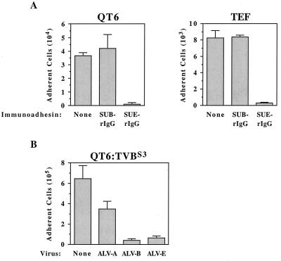
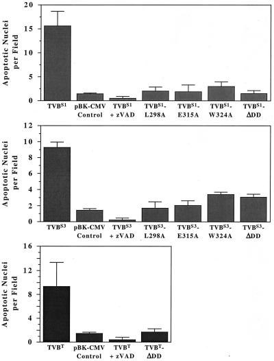
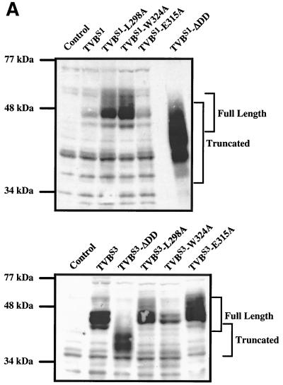
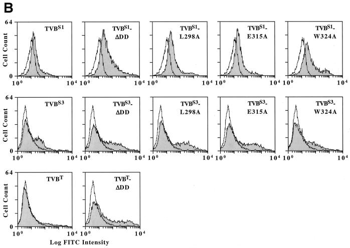
The identification of TVB(S3), a cellular receptor for the cytopathic subgroups B and D of avian leukosis virus (ALV-B and ALV-D), as a tumor necrosis factor receptor-related death receptor with a cytoplasmic death domain, provides a compelling argument that viral Env-receptor interactions are linked to cell death (4). However, other TVB proteins have been described that appear to have similar death domains but are cellular receptors for the noncytopathic subgroup E of ALV (ALV-E): TVB(T), a turkey subgroup E-specific ALV receptor, and TVB(S1), a chicken receptor for subgroups B, D, and E ALV. To begin to understand the role of TVB receptors in the cytopathic effects associated with infection by specific ALV subgroups, we asked whether binding of a soluble ALV-E surface envelope protein (SU) to its receptor can lead to cell death. Here we report that ALV-E SU-receptor interactions can induce apoptosis in quail or turkey cells. We also show directly that TVB(S1) and TVB(T) are functional death receptors that can trigger cell death by apoptosis via a mechanism involving their cytoplasmic death domains and activation of the caspase pathway. These data demonstrate that ALV-B and ALV-E use functional death receptors to enter cells, and it remains to be determined why only subgroups B and D viral infections lead specifically to cell death.


Figures

FIG. 1
FIG. 1
(A) Avian cells that express subgroup E viral receptors die specifically when incubated with an ALV-E SU-IgG fusion protein. Quail QT6-cells and primary TEFs were incubated in medium containing cycloheximide and either no immunoadhesin (None), SUB-rIgG, or SUE-rIgG. The numbers of adherent cells that survived this treatment are shown from a representative experiment performed in triplicate with the standard deviations indicated. (B) ALV-B- and ALV-E-infected cells are induced to die specifically in the presence of cycloheximide. Transfected QT6 cells expressing TVBS3 (QT6:TVBS3) were infected with subgroup A, B, and E ALV vectors and then incubated with cycloheximide. The average numbers of adherent cells surviving this treatment are shown from a representative experiment that was performed in triplicate, and the standard deviations of the data are indicated with error bars.
FIG. 2
FIG. 2
TVBS1, TVBS3, and TVBT proteins induce apoptosis via the caspase pathway by a mechanism involving their cytoplasmic death domains. Human 293 cells were transfected with plasmid DNA encoding wild-type and mutant TVB proteins or instead with a control plasmid pBK-CMV (Stratagene). Cells were incubated with or without the caspase inhibitor zVAD-fmk (zVAD). The numbers of apoptotic nuclei associated with each cell population was determined by fluorescence microscopy after Hoechst staining by using a Nikon TE-200 microscope. Ten fields of each cell population were studied. The experiments were performed in triplicate, and the standard deviations of the data obtained are shown.
FIG. 3
FIG. 3
Wild-type and mutant TVB proteins are expressed in transfected human 293 cells. (A) Human 293 cells were transfected with plasmids encoding wild-type or mutant TVB receptors or, for control purposes, with pBK vector (Stratagene). Protein lysates from these cells were subjected to immunoblotting using SUB-rIgG and an HRP-coupled secondary antibody as binding probes. The positions of the full-length and truncated (ΔDD) receptors are indicated. (B) Human 293 cells expressing wild-type and mutant TVB receptors were incubated with SUB-rIgG (TVBS1-based and TVBS3-based constructs) or with SUE-rIgG (TVBT-based constructs) and then with a fluoresceinated secondary antibody (1, 26). The cells were then analyzed by flow cytometry. Open histograms represent data from cells that were not transfected, and the shaded histograms represent data from cells expressing the different TVB proteins as indicated.
FIG. 3
FIG. 3
Wild-type and mutant TVB proteins are expressed in transfected human 293 cells. (A) Human 293 cells were transfected with plasmids encoding wild-type or mutant TVB receptors or, for control purposes, with pBK vector (Stratagene). Protein lysates from these cells were subjected to immunoblotting using SUB-rIgG and an HRP-coupled secondary antibody as binding probes. The positions of the full-length and truncated (ΔDD) receptors are indicated. (B) Human 293 cells expressing wild-type and mutant TVB receptors were incubated with SUB-rIgG (TVBS1-based and TVBS3-based constructs) or with SUE-rIgG (TVBT-based constructs) and then with a fluoresceinated secondary antibody (1, 26). The cells were then analyzed by flow cytometry. Open histograms represent data from cells that were not transfected, and the shaded histograms represent data from cells expressing the different TVB proteins as indicated.

References

    1. Adkins H B, Brojatsch J, Naughton J, Rolls M M, Pesola J M, Young J A T. Identification of a cellular receptor for subgroup E avian leukosis virus. Proc Natl Acad Sci USA. 1997;94:11617–11622. - PMC - PubMed
    1. Adkins H B, Brojatsch J, Young J A T. Identification and characterization of a shared TNFR-related receptor for subgroups B, D, and E avian leukosis viruses reveals cysteine residues required specifically for subgroup E viral entry. J Virol. 2000;74:3572–3578. - PMC - PubMed
    1. Bergeron L, Sodroski J. Dissociation of unintegrated viral DNA accumulation from single-cell lysis induced by human immunodeficiency virus type 1. J Virol. 1992;66:5777–5787. - PMC - PubMed
    1. Brojatsch J, Naughton J, Rolls M M, Zingler K, Young J A T. CAR1, a TNFR-related protein, is a cellular receptor for cytopathic avian leukosis-sarcoma viruses and mediates apoptosis. Cell. 1996;87:845–855. - PubMed
    1. Chirmule N, Pahwa S. Envelope glycoproteins of human immunodeficiency virus type 1: profound influences on immune functions. Microbiol Rev. 1996;60:386–406. - PMC - PubMed

Publication types

Substances

LinkOut - more resources