. 2000 Dec;74(24):11495-503.
doi: 10.1128/jvi.74.24.11495-11503.2000.

Enrichment of immediate-early 1 (m123/pp89) peptide-specific CD8 T cells in a pulmonary CD62L(lo) memory-effector cell pool during latent murine cytomegalovirus infection of the lungs

Affiliations

Enrichment of immediate-early 1 (m123/pp89) peptide-specific CD8 T cells in a pulmonary CD62L(lo) memory-effector cell pool during latent murine cytomegalovirus infection of the lungs

R Holtappels et al. J Virol. 2000 Dec.

Abstract

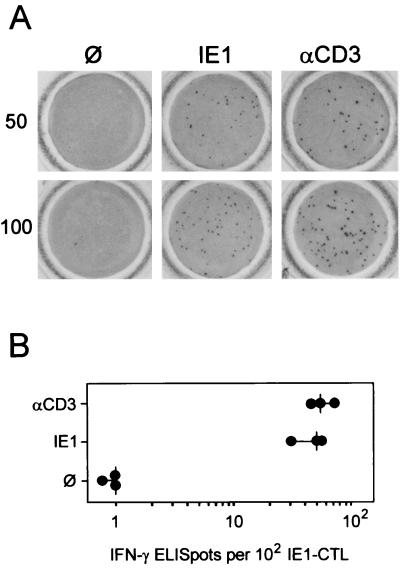
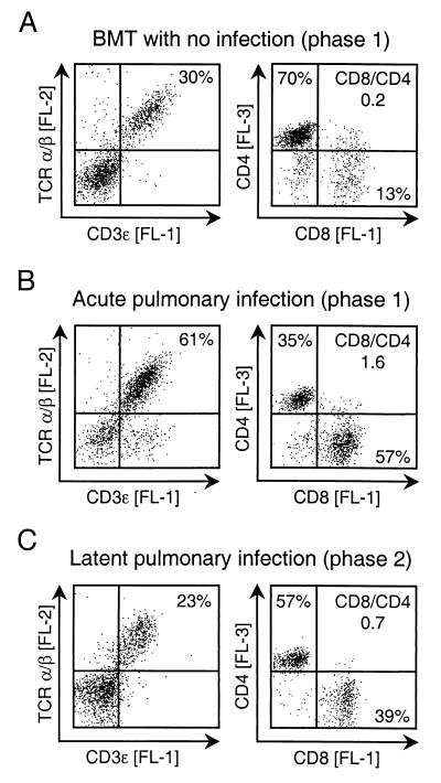
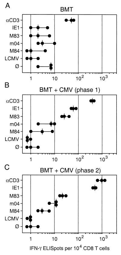
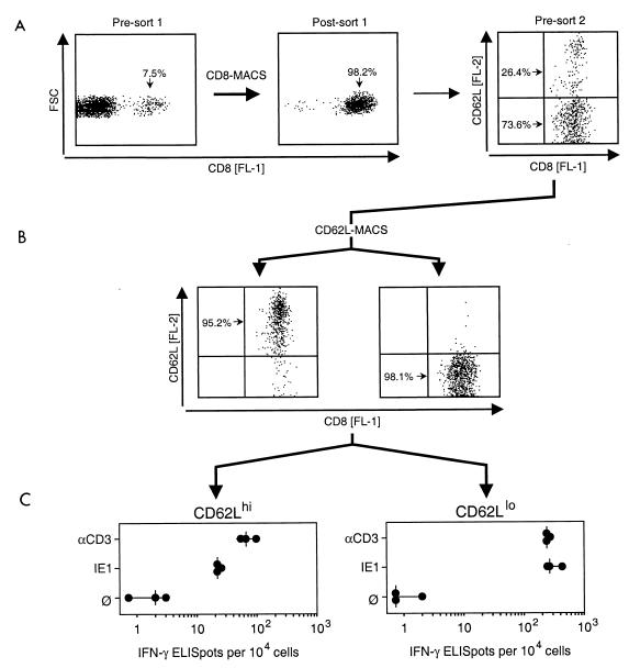
Interstitial cytomegalovirus (CMV) pneumonia is a clinically relevant complication in recipients of bone marrow transplantation (BMT). Recent data for a model of experimental syngeneic BMT and concomitant infection of BALB/c mice with murine CMV (mCMV) have documented the persistence of tissue-resident CD8 T cells after clearance of productive infection of the lungs (J. Podlech, R. Holtappels, M.-F. Pahl-Seibert, H.-P. Steffens, and M. J. Reddehase, J. Virol. 74:7496-7507, 2000). It was proposed that these cells represent antiviral "standby" memory cells whose functional role might be to help prevent reactivation of latent virus. The pool of pulmonary CD8 T cells was composed of two subsets defined by the T-cell activation marker L-selectin (CD62L): a CD62L(hi) subset of quiescent memory cells, and a CD62L(lo) subset of recently resensitized memory-effector cells. In this study, we have continued this line of investigation by quantitating CD8 T cells specific for the three currently published antigenic peptides of mCMV: peptide YPHFMPTNL processed from the immediate-early protein IE1 (pp89), and peptides YGPSLYRRF and AYAGLFTPL, derived from the early proteins m04 (gp34) and M84 (p65), respectively. IE1-specific CD8 T cells dominated in acute-phase pulmonary infiltrates and were selectively enriched in latently infected lungs. Notably, most IE1-specific CD8 T cells were found to belong to the CD62L(lo) subset representing memory-effector cells. This finding is in accordance with the interpretation that IE1-specific CD8 T cells are frequently resensitized during latent infection of the lungs and may thus be involved in the maintenance of mCMV latency.


Figures

FIG. 1
FIG. 1
Comparison between peptide-specific and CD3ɛ-redirected ELISPOT assays. Assays were performed with an IE1 (YPHFMPTNL) peptide-specific CTLL as effector cells and P815-B7 cells as the stimulating target cells. (A) Photodocumentation of ELISPOT microwell membranes for 50 and 100 IE1-CTL seeded. Shown is one of triplicate assay membranes after staining of bound IFN-γ. ∅, P815-B7 cells with no peptide added; IE1, P815-B7 cells pulsed with a saturating (10−8 M) dose of IE1 peptide; αCD3, P815-B7 cells loaded via their Fc receptors with anti-CD3ɛ MAb. (B) ELISPOT plot showing the number of spots counted. Each dot represents the result for one set of the triplicate assay cultures. The vertical dash indicates the median value.
FIG. 2
FIG. 2
Quantitative analysis of T-cell subsets in pulmonary infiltrates. The proportion of T cells among pulmonary infiltrate lymphocytes was determined by setting a lymphocyte gate in the FSC-vs-SSC plot (not shown) followed by two-color cytofluorometric analysis of the cell surface expression of CD3ɛ [FL-1] and TCR α/β [FL-2]. The proportion of CD8 T cells and CD4 T cells was determined by three-color cytofluorometric analysis of the expression of CD8 [FL-1], TCR α/β [FL-2], and CD4 [FL-3]. An electronic gate was set on positive FL-2 to restrict the analysis of CD8 and CD4 expression to α/β T lymphocytes. Shown are two-dimensional dot plots for 10,000 gated cells with 2,500 dots displayed. Quadrants were defined by isotype controls. Percentages of populations of interest are indicated in the appropriate quadrants. CD8/CD4, ratio of CD8 T cells to CD4 T cells. (A) Analysis of pulmonary leukocytes isolated at 4 weeks (phase 1) after BMT performed with no infection. (B) Analysis of pulmonary leukocytes isolated during viral replication in the lungs at 4 weeks (phase 1) after BMT and simultaneous infection with mCMV. (C) Analysis of pulmonary leukocytes isolated after resolution of productive infection at 3 months (phase 2), i.e., during viral latency in the lungs.
FIG. 3
FIG. 3
Frequencies of IFN-γ-secreting effector T cells in pulmonary infiltrates. Effector cells in the ELISPOT assays were immunomagnetically purified CD8 T cells derived from pulmonary infiltrates. (A) Infiltrates isolated at 4 weeks (phase 1) after BMT with no infection. (B) Infiltrates isolated at 4 weeks (phase 1) during acute infection of the lungs. (C) Infiltrates isolated at 3 months during latent infection of the lungs. The CD3ɛ-redirected ELISPOT assay (Fig. 1) was used to determine the frequency of effector cells irrespective of their antigen specificity. Negative controls included the presentation of an unrelated peptide (LCMV NP aa 118 to 126) by the Ld molecule of P815-B7 cells as well as omission of peptide (∅). Specific peptides of mCMV included IE1 (aa 168 to 176) presented by Ld, M83 presented by Ld, m04 (aa 243 to 251) presented by Dd, and M84 (aa 297 to 305) presented by Kd. Dots represent results from individual assay cultures. Vertical dashes indicate median values.
FIG. 4
FIG. 4
Localization of IFN-γ-secreting effector cells in CD8 T-cell subsets defined by expression of the activation marker CD62L. Interstitial leukocytes were isolated from latently infected lungs at 3 months after BMT and primary mCMV infection. (A) CD8 T cells were enriched to almost purity by positive immunomagnetic cell sorting (CD8-MACS, sort 1) and analyzed for cell surface expression of CD8 [FL-1] and L-selectin CD62L [FL-2]. FSC, forward scatter representing cell size. The two-dimensional presort 2 dot plot shows 2,500 cells. (B) Cytofluorometric reanalysis of CD8 T cells after immunomagnetic sorting into CD62Lhi and CD62Llo subsets (CD62L-MACS, sort 2). Analyses were performed for 2,500 sorted cells, with all cells displayed as dots. (C) Frequencies of IFN-γ-secreting effector cells determined by CD3ɛ-redirected and IE1 (YPHFMPTNL) peptide-specific ELISPOT assays. ∅, P815-B7 target cells with no peptide added. Dots represent results from individual assay cultures. Vertical dashes indicate median values.

References

    1. Alterio de Goss M, Holtappels R, Steffens H-P, Podlech J, Angele P, Dreher L, Thomas D, Reddehase M J. Control of cytomegalovirus in bone marrow transplantation chimeras lacking the prevailing antigen-presenting molecule in recipient tissues rests primarily on recipient-derived CD8 T cells. J Virol. 1998;72:7733–7744. - PMC - PubMed
    1. Angulo A, Ghazal P, Messerle M. The major immediate-early gene ie3 of mouse cytomegalovirus is essential for viral growth. J Virol. 2000;74:11129–11136. - PMC - PubMed
    1. Ashton-Rickardt P G, Opferman J T. Memory T lymphocytes. Cell Mol Life Sci. 1999;56:69–77. - PMC - PubMed
    1. Azuma M, Cayabyab M, Buck D, Philipps J H, Lanier L L. CD28 interaction with B7 costimulates primary allogeneic proliferative responses and cytotoxicity mediated by small, resting T lymphocytes. J Exp Med. 1992;175:353–360. - PMC - PubMed
    1. Bachmann M F, Kundig T M, Hengartner H, Zinkernagel R M. Protection against immunopathological consequences of a viral infection by activated but not resting cytotoxic T cells: T cell memory without “memory T cells”? Proc Natl Acad Sci USA. 1997;94:640–645. - PMC - PubMed

Publication types

Substances