. 2000 Dec;74(24):11608-18.
doi: 10.1128/jvi.74.24.11608-11618.2000.

A null mutation in the UL36 gene of herpes simplex virus type 1 results in accumulation of unenveloped DNA-filled capsids in the cytoplasm of infected cells

Affiliations

A null mutation in the UL36 gene of herpes simplex virus type 1 results in accumulation of unenveloped DNA-filled capsids in the cytoplasm of infected cells

P J Desai. J Virol. 2000 Dec.

Abstract

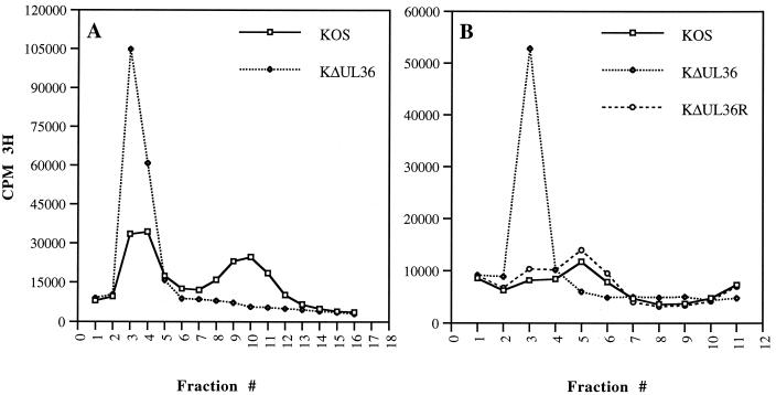
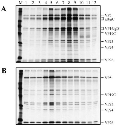
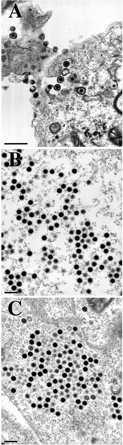
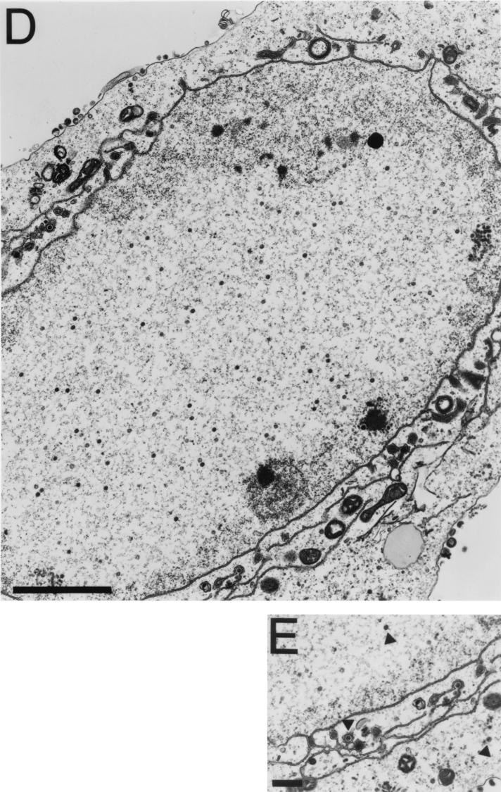
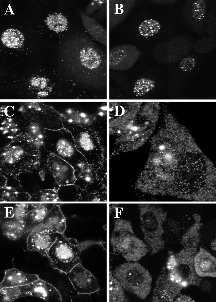
The UL36 open reading frame (ORF) encodes the largest herpes simplex virus type 1 (HSV-1) protein, a 270-kDa polypeptide designated VP1/2, which is also a component of the virion tegument. A null mutation was generated in the UL36 gene to elucidate its role in the virus life cycle. Since the UL36 gene specifies an essential function, complementing cell lines transformed for sequences encoding the UL36 ORF were made. A mutant virus, designated KDeltaUL36, that encodes a null mutation in the UL36 gene was isolated and propagated in these cell lines. When noncomplementing cells infected with KDeltaUL36 were analyzed, both terminal genomic DNA fragments and DNA-containing capsids (C capsids) were detected; therefore, UL36 is not required for cleavage or packaging of DNA. Sedimentation analysis of lysates from mutant-infected cells revealed the presence of particles that have the physical characteristics of C capsids. In agreement with this, polypeptide profiles of the mutant particles revealed an absence of the major envelope and tegument components. Ultrastructural analysis revealed the presence of numerous unenveloped DNA containing capsids in the cytoplasm of KDeltaUL36-infected cells. The UL36 mutant particles were tagged with the VP26-green fluorescent protein marker, and their movement was monitored in living cells. In KDeltaUL36-infected cells, extensive particulate fluorescence corresponding to the capsid particles was observed throughout the cytosol. Accumulation of fluorescence at the plasma membrane which indicated maturation and egress of virions was observed in wild-type-infected cells but was absent in KDeltaUL36-infected cells. In the absence of UL36 function, DNA-filled capsids are produced; these capsids enter the cytosol after traversing the nuclear envelope and do not mature into enveloped virus. The maturation and egress of the UL36 mutant particles are abrogated, possibly due to a late function of this complex polypeptide, i.e., to target capsids to the correct maturation pathway.


Figures

FIG. 1
FIG. 1
Schematic representation of the BglII D region of the KOS genome. The BglII D (25.9-kb) fragment encodes UL31 to UL38 (29). The direction of the UL36 transcript is indicated below the ORF. The UL36 ORF starts at genome nucleotide 80543 and stops at 71051. Three cell lines that express UL36 were made: B80, which was transformed with the BglII D fragment (pKBGD); XS13, transformed with the XbaI-to-SnaB1 fragment (pKXSB); and HS30, transformed with the HpaI-to-SnaB1 fragment containing a deletion in the UL35 gene (pKUL36). The deletion in pKΔUL36 spans from the KpnI to EcoRV sites in UL36. Relevant restriction enzyme sites and genome nucleotide numbers (29) in parentheses are indicated at the top.
FIG. 2
FIG. 2
Infected cell polypeptide synthesis in KΔUL36-infected cells. Vero cells (106 in 35-mm-diameter dishes) were infected with KOS (lane 2) and KΔUL36 (lane 3) at an MOI of 10 PFU/cell or mock infected (lane 1). The infected cells were metabolically labeled with [35S]methionine from 9 to 24 h postinfection. Cells were solubilized in Laemmli sample buffer, and the proteins were analyzed by SDS-PAGE (9% acrylamide). Protein standards (lane M) correspond to 220, 97.4, 66, and 46 kDa (the 97.4-kDa marker migrates as a doublet in our gels). The closed circle indicates the position of the UL36 protein in KOS lysates.
FIG. 3
FIG. 3
Cleavage of viral genomic DNA in KΔUL36-infected cells. Vero (lanes 1 and 2) and HS30 (lanes 3 and 4) cells (106 in 35-mm-diameter dishes) were infected with KOS (lanes 1 and 3) and KΔUL36 (lanes 2 and 4) at an MOI of 10 PFU/cell. Infected cell DNA was prepared 24 h postinfection, and 2 μg was digested with BamHI. The restriction fragments were analyzed by Southern blot hybridization using a 32P-labeled DNA fragment corresponding to the BamHI K junction fragment. The BamHI K junction fragment and terminal Q and S fragments are indicated at the right; shown below the blot is a schematic representation of the HSV-1 genome and locations of the BamHI junction and terminal fragments.
FIG. 4
FIG. 4
Capsid formation in KΔUL36-infected cells. Vero cell monolayers (107 cells) were infected with KOS (A) and KΔUL36 (B) at an MOI of 10 PFU/cell and labeled with [35S]methionine from 8 to 24 h postinfection. Nuclear extracts were prepared and layered onto 20 to 50% sucrose gradients. Fractions collected after sedimentation were TCA precipitated, and the proteins in the fractions were resolved by SDS-PAGE (17% acrylamide). Direction of sedimentation was from right to left. The positions of capsid proteins are indicated at the left for KOS; the positions at which A, B, and C capsids sediment are indicated at the bottom.
FIG. 5
FIG. 5
Particle formation in KΔUL36-infected cells. HEL (2 × 10⁷) (A) and Vero (107) (B) cells infected with KOS (A and B), KΔUL36 (A and B), and KΔUL36R (B) at an MOI of 10 PFU/cell were metabolically labeled with [3H]thymidine from 8 to 24 h postinfection. Intracellular virus was pelleted, loaded onto 20 to 50% sucrose gradients, and subjected to rate velocity sedimentation. The radioactivity present in the fractions collected was determined by liquid scintillation. Fraction 1 corresponds to the bottom of the tube.
FIG. 6
FIG. 6
SDS-PAGE analysis of KΔUL36 particles. HEL cells (2 × 10⁷) infected with KOS and KΔUL36 at an MOI of 10 PFU/cell were labeled with [35S]methionine from 8 to 24 h after infection. Cell lysates were sedimented through 20 to 50% sucrose gradients, the light-scattering bands were harvested by side puncture, and the particles were pelleted and again sedimented through 20 to 50% sucrose gradients. Fractions collected after sedimentation were TCA precipitated, and the proteins in the fractions were resolved by SDS-PAGE (17% acrylamide). Fraction 1 corresponds to the bottom of the tube. Relevant virion proteins are indicated at the right; protein standards (lane M) correspond to 220, 97.4, 66, 46, 30, and 14.3 kDa (indicated by the closed circles).
FIG. 7
FIG. 7
TEM analysis of KΔUL36-infected cells. Vero cells infected with KOS (A) and KΔUL36 (B to E) at an MOI of 10 PFU/cell were fixed at 16 h postinfection, thin sectioned, and processed for TEM. Magnifications were ×27,500 (A), ×37,000 (B and C), ×13,600 (D), and ×20,500 (E). Bars = 500 nm (A and E), 200 nm (B and C), and 2,000 nm (D).
FIG. 7
FIG. 7
TEM analysis of KΔUL36-infected cells. Vero cells infected with KOS (A) and KΔUL36 (B to E) at an MOI of 10 PFU/cell were fixed at 16 h postinfection, thin sectioned, and processed for TEM. Magnifications were ×27,500 (A), ×37,000 (B and C), ×13,600 (D), and ×20,500 (E). Bars = 500 nm (A and E), 200 nm (B and C), and 2,000 nm (D).
FIG. 8
FIG. 8
Analysis in living cells of the replication of GFP-tagged KΔUL36. Cells infected with K26GFP (A, C, and E) and KΔUL36-GFP (B, D, and F) at an MOI of 10 PFU/cell were visualized live in a confocal microscope at 8 (A and B), 12 (C and D), and 18 (E and F) h after infection. Magnification was ×60 (A, B, C, E, and F) or ×100 (D).

References

    1. Ace C I, Dalrymple M A, Ramsay F H, Preston V G, Preston C M. Mutational analysis of the herpes simplex virus type 1 trans-inducing factor Vmw65. J Gen Virol. 1988;69:2595–2605. - PubMed
    1. Baines J D, Roizman B. The UL11 gene of herpes simplex virus type 1 encodes a function that facilitates nucleocapsid envelopment and egress from cells. J Virol. 1992;66:5168–5174. - PMC - PubMed
    1. Batterson W, Furlong D, Roizman B. Molecular genetics of herpes simplex virus. VIII. Further characterization of a temperature-sensitive mutant defective in release of viral DNA and in other stages of the viral reproductive cycle. J Virol. 1983;45:397–407. - PMC - PubMed
    1. Batterson W, Roizman B. Characterization of the herpes simplex virion-associated factor responsible for the induction of α genes. J Virol. 1983;46:371–377. - PMC - PubMed
    1. Browne H B, Bell S, Minson T, Wilson D W. An endoplasmic reticulum-retained herpes simplex virus glycoportein H is absent from secreted virions: evidence for reenvelopment during egress. J Virol. 1996;70:4311–4316. - PMC - PubMed

Publication types