. 2000 Dec;74(24):11690-6.
doi: 10.1128/jvi.74.24.11690-11696.2000.

Profound protection against respiratory challenge with a lethal H7N7 influenza A virus by increasing the magnitude of CD8(+) T-cell memory

Affiliations

Profound protection against respiratory challenge with a lethal H7N7 influenza A virus by increasing the magnitude of CD8(+) T-cell memory

J P Christensen et al. J Virol. 2000 Dec.

Abstract

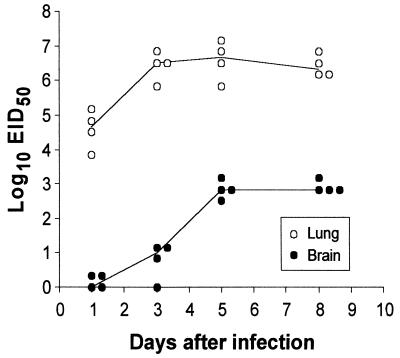
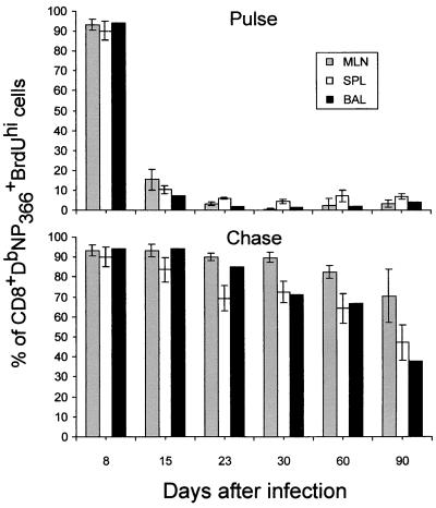
The recall of CD8(+) T-cell memory established by infecting H-2(b) mice with an H1N1 influenza A virus provided a measure of protection against an extremely virulent H7N7 virus. The numbers of CD8(+) effector and memory T cells specific for the shared, immunodominant D(b)NP(366) epitope were greatly increased subsequent to the H7N7 challenge, and though lung titers remained as high as those in naive controls for 5 days or more, the virus was cleared more rapidly. Expanding the CD8(+) memory T-cell pool (<0.5 to >10%) by sequential priming with two different influenza A viruses (H3N2-->H1N1) gave much better protection. Though the H7N7 virus initially grew to equivalent titers in the lungs of naive and double-primed mice, the replicative phase was substantially controlled within 3 days. This tertiary H7N7 challenge caused little increase in the magnitude of the CD8(+) D(b)NP(366)(+) T-cell pool, and only a portion of the memory population in the lymphoid tissue could be shown to proliferate. The great majority of the CD8(+) D(b)NP(366)(+) set that localized to the infected respiratory tract had, however, cycled at least once, though recent cell division was shown not to be a prerequisite for T-cell extravasation. The selective induction of CD8(+) T-cell memory can thus greatly limit the damage caused by a virulent influenza A virus, with the extent of protection being directly related to the number of available responders. Furthermore, a large pool of CD8(+) memory T cells may be only partially utilized to deal with a potentially lethal influenza infection.


Figures

FIG. 1
FIG. 1
Virus titers in lung and brain homogenates after respiratory exposure to the H7N7 influenza virus. The B6 mice were infected i.n. with 400 EID50 of the H7N7 virus, and blood, spleen, lung, and brain samples were taken for virus recovery. No virus was detected in the blood or spleen. The data are cumulative from two experiments and show results for individual mice. A further study established that there is indeed an eclipse phase for the H7N7 virus. Essentially no infectious virus was detected in lung homogenates from five mice sampled (100.2±0.3) 4 h after i.n. challenge with 400 EID50, though new virus was emerging by 8 h (102.0±0.3).
FIG. 2
FIG. 2
Comparison of H7N7 virus replication in naive and single- and double-primed mice. Age-matched B6 female mice were infected i.n. with 1,000 EID50 of the H7N7 virus after either no prior experience with an influenza A virus (1°), i.p. infection with an H1N1 virus (2°), or further i.n. challenge of the H1N1-primed mice with an H3N2 virus (3°). All mice were rested for at least 1 month between each infection. The lungs and brains were removed at intervals for virus titration and the analysis of virus-specific CD8+ T-cell numbers (Fig. 3). The data are expressed as the means ± the standard deviations for five mice per group. The remaining mice in the 1° group had succumbed by day 13 after infection.
FIG. 3
FIG. 3
Total number of CD8+ DbNP366+ T cells after primary, secondary, and tertiary influenza infection. This analysis utilized the same mice that were assayed for Fig. 2. The MLN and spleen (SPL) samples were analyzed from five individuals, while the BAL samples were pooled. The tetramer staining results are expressed as mean values (BAL) or as the means ± the standard deviations.
FIG. 4
FIG. 4
Visualization of CD8+ DbNP366+-specific populations recovered from different anatomical sites 7 days after i.n. challenge of H3N2→H1N1-primed mice with the H7N7 virus. The CSF samples were obtained from the cisterna magna of anesthetized, exsanguinated mice.
FIG. 5
FIG. 5
Acute proliferation and long-term cycling of CD8+ DbNP366+ T cells in secondarily stimulated (H3N2→H1N1) mice. The mice in the pulse analysis were given BrdU in drinking water for 8 days prior to sampling. Those in the chase study were fed BrdU from day 0 to 8 following secondary challenge. The results, which show only the values for the BrdUhi subset, are the means ± the standard deviations for groups of five mice.
FIG. 6
FIG. 6
Cycling characteristics of the CD8+ DbNP366+ set recovered 8 days after i.n. challenge of double-primed (H3N2→H1N1) mice with the H7N7 virus or with an influenza B virus. All mice were injected i.p. with the H1N1 virus, given the H3N2 virus i.n. 1 month later, and rested for a further 6 months before i.n. challenge with the H7N7 virus or the B/HK (FluB) virus. The mice in the pulse experiment (left half) were given BrdU in the drinking water for the 8 days after i.n. exposure to the H7N7 or B/HK virus, while those in the chase study (right half) had been fed BrdU 6 months previously, at the time of the secondary H3N2 stimulation. Cycling through the pulse analysis is thus characterized by BrdU incorporation (BrdUhi) and through the chase study by the loss of BrdU (BrdUlo).

References

    1. Allan W, Tabi Z, Cleary A, Doherty P C. Cellular events in the lymph node and lung of mice with influenza. Consequences of depleting CD4+ T cells. J Immunol. 1990;144:3980–3986. - PubMed
    1. Andrew M E, Coupar B E. Efficacy of influenza haemagglutinin and nucleoprotein as protective antigens against influenza virus infection in mice. Scand J Immunol. 1988;28:81–85. - PubMed
    1. Banchereau J. Dendritic cells: therapeutic potentials. Transfus Sci. 1997;18:313–326. - PubMed
    1. Belz G T, Xie W, Altman J D, Doherty P C. A previously unrecognized H-2Db-restricted peptide prominent in the primary influenza A virus-specific CD8+ T-cell response is much less apparent following secondary challenge. J Virol. 2000;74:3486–3493. - PMC - PubMed
    1. Bender B S, Small P A., Jr Heterotypic immune mice lose protection against influenza virus infection with senescence. J Infect Dis. 1993;168:873–880. - PubMed

Publication types