. 2000 Dec;74(24):11744-53.
doi: 10.1128/jvi.74.24.11744-11753.2000.

Wide range of viral load in healthy african green monkeys naturally infected with simian immunodeficiency virus

Affiliations

Wide range of viral load in healthy african green monkeys naturally infected with simian immunodeficiency virus

S Goldstein et al. J Virol. 2000 Dec.

Abstract

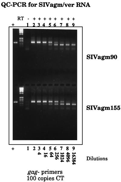
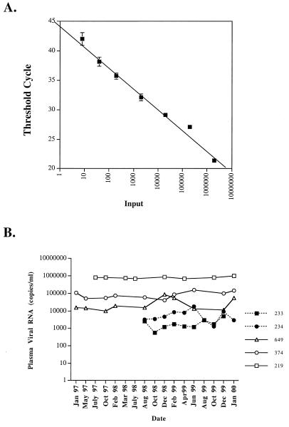
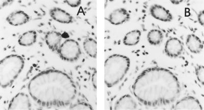
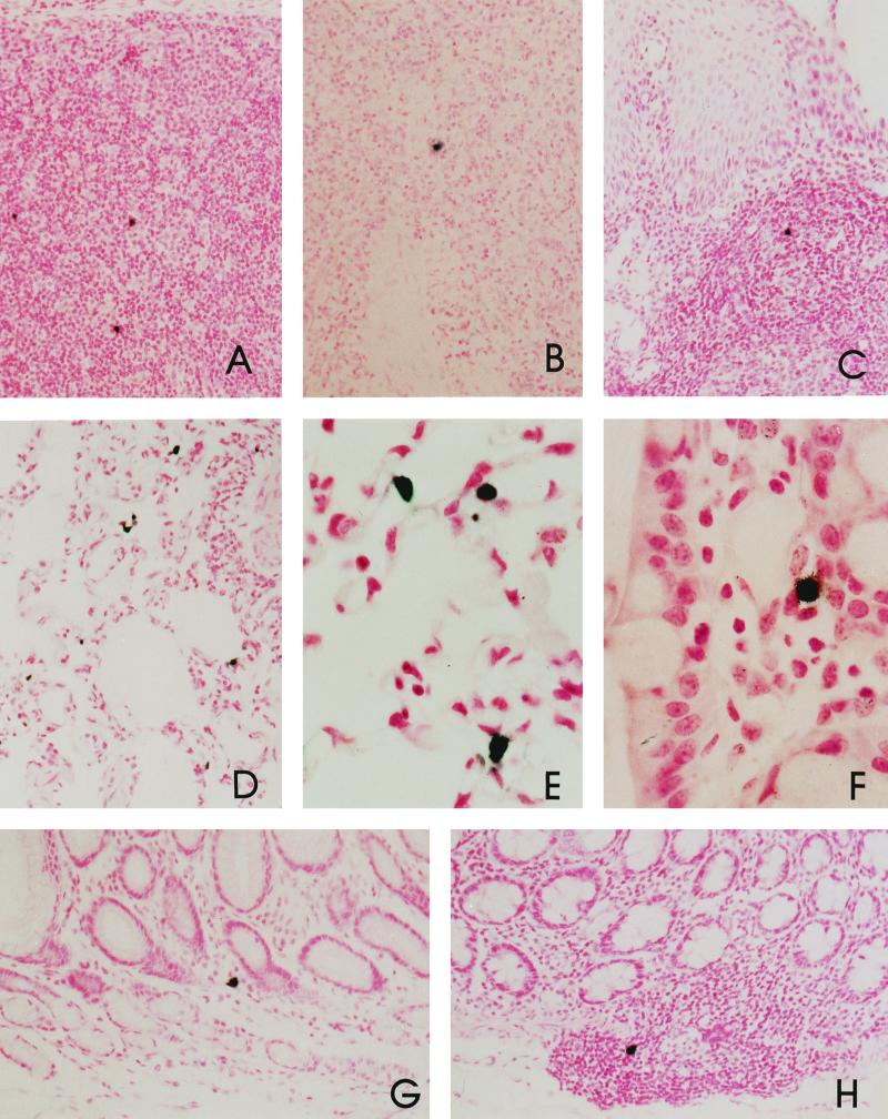
The distribution and levels of simian immunodeficiency virus (SIV) in tissues and plasma were assessed in naturally infected African green monkeys (AGM) of the vervet subspecies (Chlorocebus pygerythrus) by limiting-dilution coculture, quantitative PCR for viral DNA and RNA, and in situ hybridization for SIV expression in tissues. A wide range of SIV RNA levels in plasma was observed among these animals (<1,000 to 800,000 copies per ml), and the levels appeared to be stable over long periods of time. The relative numbers of SIV-expressing cells in tissues of two monkeys correlated with the extent of plasma viremia. SIV expression was observed in lymphoid tissues and was not associated with immunopathology. Virus-expressing cells were observed in the lamina propria and lymphoid tissue of the gastrointestinal tract, as well as within alveolar macrophages in the lung tissue of one AGM. The range of plasma viremia in naturally infected AGM was greater than that reported in naturally infected sooty mangabeys. However, the degree of viremia in some AGM was similar to that observed during progression to AIDS in human immunodeficiency virus-infected individuals. Therefore, containment of viremia is an unlikely explanation for the lack of pathogenicity of SIVagm in its natural host species, AGM.


Figures

FIG. 1
FIG. 1
Oligonucleotide primers used for quantitative PCR for viral RNA. (A) Primers used for QC-PCR assay. Shown is an alignment of portions of the gag gene of SIVagm clones from vervet monkeys compared with SIVagm9063-2, where identity is shown as a dot and nucleotide substitutions are indicated. Nucleotide positions within the genome of SIVagm9063-2 are indicated at the right. The forward (F) and reverse (R) primers used for the QC-PCR assay are shown below the sequence alignment. (B) Primers used for real-time PCR assay. Shown is an alignment of portions of the envelope gene of SIVagm clones compared with that of SIVagm9063-2, where identity is shown as a dot and nucleotide substitutions are indicated. The primers used for real-time PCR analysis are shown below the alignment, with the letter i indicating insertion of an inosine at a variable amino acid.
FIG. 2
FIG. 2
Analysis of SIVagm isolates by QC-PCR. PCR amplifications of reactions containing 100 copies of the competitive template (CT) and of serial fourfold dilution series of viral RNAs extracted from SIVagm90- and SIVagm155-infected-cell culture supernatants are shown. The plus and minus signs on the top line indicate whether RT was included in the reaction mixture. Virus was used undiluted (lane 2) or diluted 1:4 (lane 3), 1:16 (lane 4); 1:64 (lane 5), 1:256 (lane 6), 1:1,024 (lane 7), 1:4,096 (lane 8), or 1:16,384 (lane 9).
FIG. 3
FIG. 3
Analysis of SIVagm RNA levels in plasma of naturally and experimentally infected AGM using a real-time RT-PCR assay. This plot of the input control template (in vitro envelope transcript) copy number versus the threshold cycle demonstrates the assay's precision and broad dynamic range. The plotted values represent mean values ± 1 standard deviation for triplicate determinations. (B) SIV RNA levels in plasma through 3 years of chronic infection in a naturally infected vervet (AGM219). Sequential SIV RNA levels in plasma are also shown for two vervet monkeys (374 and 649) and two sabaeus monkeys (233 and 234) that were experimentally inoculated with SIVagm9063 in 1994.
FIG. 4
FIG. 4
Specificity of detection of SIV expression by ISH. SIV expression in the gastrointestinal tract of AGM90 showing the specificity of hybridization. (A) Section hybridized with the antisense SIV probe showing an SIV-expressing cell near an intestinal crypt. (B) Same field as in panel A on a serial section hybridized with the sense SIV probe demonstrating no specific hybridization.
FIG. 5
FIG. 5
Examples of SIV-expressing cells in lymphoid and nonlymphoid tissues. SIV expression in the gastrointestinal tracts of naturally infected AGM90 and AGM155 was detected by ISH. Panels: A, mesenteric lymph node of AGM90; B, spleen of AGM90; C, tonsil of AGM90; D and E, macrophages within the lung tissue of AGM90; F, ileum of AGM155; G, stomach of AGM90; H, colon of AGM90.

References

    1. Allan J S, Short M, Taylor M E, Su S, Hirsch V M, Johnson P R, Shaw G M, Hahn B H. Species-specific diversity among simian immunodeficiency viruses from African green monkeys. J Virol. 1991;65:2816–2828. - PMC - PubMed
    1. Allan J S, Kanda P, Kennedy R C, Cobb E K, Anthony M, Eichberg J W. Isolation and characterization of simian immunodeficiency viruses from two subspecies of African Green monkeys. AIDS Res Hum Retrovir. 1990;6:275–285. - PubMed
    1. Allan J S. Pathogenic properties of simian immunodeficiency viruses in nonhuman primates. Annu Rev AIDS Res. 1991;1:191–206.
    1. Baier M, Garber C, Cichutek K, Kurth R. Complete nucleotide sequence of a simian immunodeficiency virus from African green monkeys: a novel type of intragroup divergence. Virology. 1990;176:216–221. - PubMed
    1. Baier M, Werner A, Cichutek K, Garber C, Müller C, Kraus G, Ferdinand F J, Hartung S, Papas T S, Kurth R. Molecularly cloned simian immunodeficiency virus SIVagm3 is highly divergent from other SIVagm isolates and is biologically active in vitro and in vivo. J Virol. 1989;63:5119–5123. - PMC - PubMed

LinkOut - more resources