. 2000 Dec;74(24):11764-72.
doi: 10.1128/jvi.74.24.11764-11772.2000.

Development of a primary tamarin hepatocyte culture system for GB virus-B: a surrogate model for hepatitis C virus

Affiliations

Development of a primary tamarin hepatocyte culture system for GB virus-B: a surrogate model for hepatitis C virus

B Beames et al. J Virol. 2000 Dec.

Abstract

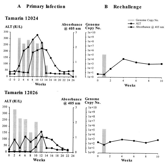
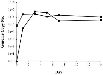
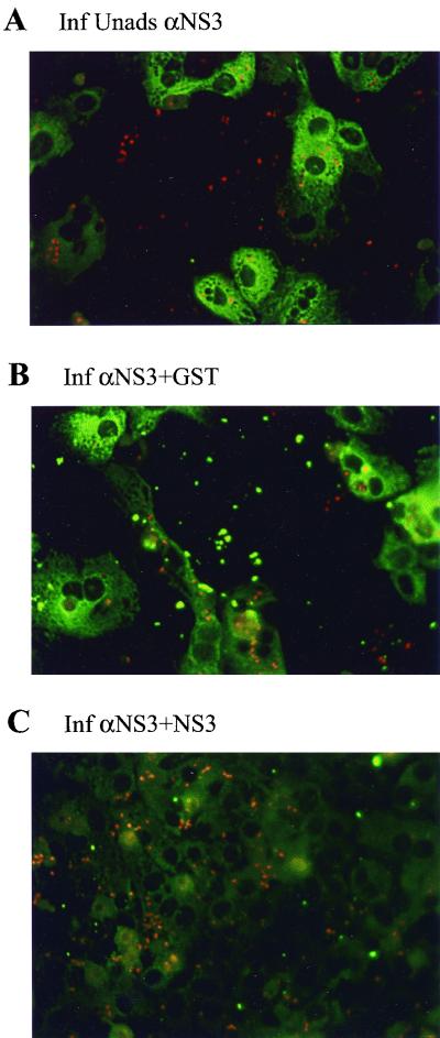
GB virus-B (GBV-B) causes an acute hepatitis in tamarins characterized by increased alanine transaminase levels that quickly return to normal as the virus is cleared. Phylogenetically, GBV-B is the closest relative to hepatitis C virus (HCV), and thus GBV-B infection of tamarins represents a powerful surrogate model system for the study of HCV. In this study, the course of infection of GBV-B in tamarins was followed using a real-time 5' exonuclease (TaqMan) reverse transcription-PCR assay to determine the level of GBV-B in the serum. Peak viremia levels exceeded 10(9) genome equivalents/ml, followed by viral clearance within 14 to 16 weeks. Rechallenge of animals that had cleared infection resulted in viremia that was limited to 1 week, suggestive of a strong protective immune response. A robust tissue culture system for GBV-B was developed using primary cultures of tamarin hepatocytes. Hepatocytes obtained from a GBV-B-infected animal maintained high levels of cell-associated viral RNA and virion secretion for 42 days of culture. In vitro infection of normal hepatocytes resulted in rapid amplification of cell-associated viral RNA and secretion of up to 10(7) genome equivalents/ml of culture supernatant. In addition, infection could be monitored by immunofluorescence staining for GBV-B nonstructural NS3 protein. This model system overcomes many of the current obstacles to HCV research, including low levels of viral replication, lack of a small primate animal model, and lack of a reproducible tissue culture system.


Figures

FIG. 1
FIG. 1
Infection profiles of GBV-B infected tamarins. (A) Progress of initial GBV-B infection in tamarins 12024 (top) and 12026 (bottom). GBV-B levels are shown in comparison to the liver damage, as measured by ALT, and rise in serum antibody titers to the GBV-B NS3 protein over the 24- to 26-week course of the experiment. (B) GBV-B levels and serum antibody titers to the NS3 protein during the course of rechallenge infection of 12024 (top) and 12026 (bottom) with a standard inoculum of GBV-B-containing 12024 tamarin serum. GBV-B TaqMan data, measured in genome equivalents per milliliter of serum, are shown as bar graphs, with line graphs representing ALT and NS3 ELISA absorbance superimposed.
FIG. 2
FIG. 2
Persistence of GBV-B replication in primary tamarin hepatocytes. GBV-B levels were monitored for 42 days in primary hepatocytes isolated from GBV-B-infected tamarin 12036. Day 0 represents RNA isolated from hepatocytes harvested on the day of isolation from the liver.
FIG. 3
FIG. 3
Growth curve of GBV-B in primary tamarin hepatocytes. Tamarin hepatocytes were inoculated with GBV-B-containing plasma, using an adsorption period of 6 h. Cultures were washed extensively to remove unadsorbed virus and then harvested in duplicate at various times over a 14-day period. Time zero (immediately after washing away the inoculum) represents the level of virus attached or internalized by the cells. Cell-associated (squares) or secreted (circles) GBV-B RNA was quantified by TaqMan RT-PCR and expressed as genome equivalents per microgram of cellular RNA or milliliter of culture medium, respectively.
FIG. 4
FIG. 4
Estimation of doubling time for GBV-B in primary tamarin hepatocytes. Tamarin hepatocytes were inoculated with GBV-B-containing plasma using a shortened adsorption period (1 h). Cultures were washed extensively to remove unadsorbed virus and harvested in duplicate at various times over a 48-h period. Time (immediately after washing away the inoculum) represents the level of virus attached or internalized by the cells. Cell-associated GBV-B RNA was quantified by TaqMan RT-PCR.
FIG. 5
FIG. 5
Immunofluorescence staining for NS3 protein. Hepatocytes grown on glass coverslips were inoculated with GBV-B 3 days postplating and harvested 3 days postinfection unless noted otherwise. GBV-B NS3 protein was detected by immunofluorescence using a rabbit anti-GST-NS3 antiserum and goat anti-rabbit IgG-fluorescein. (A) Infected hepatocytes stained with anti-NS3 (magnification, ×100); (B) uninfected hepatocytes stained with anti-NS3 (×100); (C) infected hepatocytes stained with normal rabbit (prebleed; ×100); (D) lower magnification of infected hepatocytes stained with anti-NS3 (×50); (E) hepatocytes infected with a low multiplicity and harvested 21 days p.i. (×100); (F) higher magnification of infected hepatocytes stained with anti-NS3 (×200).
FIG. 6
FIG. 6
Specificity of rabbit anti-GST-NS3 antiserum in immunofluorescence staining for NS3 protein. Hepatocytes were inoculated with GBV-B and harvested 3 days p.i. Cells were stained for NS3 protein using rabbit anti-GST-NS3 antiserum (A). To demonstrate specificity, the antiserum was adsorbed for 16 h at 4°C with 5 μg of purified GST (B) or NS3 (C) protein prior to being used for immunofluorescence staining. Adsorption with GST had no effect on staining, while adsorption with NS3 eliminated staining.

References

    1. Alter H, Nakatsuji Y, Melpolder J, Wages J, Jr, Wesley R, Shih J W K, Kim J P. The incidence of transfusion-associated hepatitis G virus infection and its relation to liver disease. N Engl J Med. 1997;336:747–754. - PubMed
    1. Bassett S E, Brasky K M, Lanford R E. Analysis of hepatitis C virus-inoculated chimpanzees reveals unexpected clinical profiles. J Virol. 1998;72:2589–2599. - PMC - PubMed
    1. Beard M R, Abell G, Honda M, Carroll A, Gartland M, Clarke B, Suzuki K, Lanford R, Sangar D V, Lemon S M. An infectious molecular clone of a Japanese genotype 1b hepatitis C virus. Hepatology. 1999;30:316–324. - PubMed
    1. Bronowicki J P, Vetter D, Uhl G, Hudziak H, Uhrlacher A, Vetter J M, Doffoel M. Lymphocyte reactivity to hepatitis C virus (HCV) antigens shows evidence for exposure to HCV in HCV-seronegative spouses of HCV-infected patients. J Infect Dis. 1997;176:518–522. - PubMed
    1. Bukh J, Apgar C L. Five new or recently discovered (GBV-A) virus species are indigenous to New World monkeys and may constitute a separate genus of the Flaviviridae. Virology. 1997;229:429–436. - PubMed

Publication types

LinkOut - more resources