. 2000 Dec;74(24):11792-9.
doi: 10.1128/jvi.74.24.11792-11799.2000.

Mechanism of interference mediated by human parainfluenza virus type 3 infection

Affiliations

Mechanism of interference mediated by human parainfluenza virus type 3 infection

M A Horga et al. J Virol. 2000 Dec.

Abstract

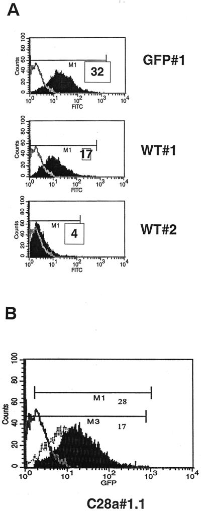
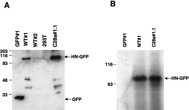
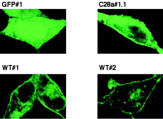
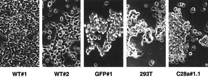
Viral interference is characterized by the resistance of infected cells to infection by a challenge virus. Mechanisms of viral interference have not been characterized for human parainfluenza virus type 3 (HPF3), and the possible role of the neuraminidase (receptor-destroying) enzyme of the hemagglutinin-neuraminidase (HN) glycoprotein has not been assessed. To determine whether continual HN expression results in depletion of the viral receptors and thus prevents entry and cell fusion, we tested whether cells expressing wild-type HPF3 HN are resistant to viral infection. Stable expression of wild-type HN-green fluorescent protein (GFP) on cell membranes in different amounts allowed us to establish a correlation between the level of HN expression, the level of neuraminidase activity, and the level of protection from HPF3 infection. Cells with the highest levels of HN expression and neuraminidase activity on the cell surface were most resistant to infection by HPF3. To determine whether this resistance is attributable to the viral neuraminidase, we used a cloned variant HPF3 HN that has two amino acid alterations in HN leading to the loss of detectable neuraminidase activity. Cells expressing the neuraminidase-deficient variant HN-GFP were not protected from infection, despite expressing HN on their surface at levels even higher than the wild-type cell clones. Our results demonstrate that the HPF3 HN-mediated interference effect can be attributed to the presence of an active neuraminidase enzyme activity and provide the first definitive evidence that the mechanism for attachment interference by a paramyxovirus is attributable to the viral neuraminidase.


Figures

FIG. 1
FIG. 1
FACS analysis of clonal cell lines expressing HN-GFP. (A) FACS analysis of WT#1 and WT#2 cell lines. The x and y axes of the histograms represent green fluorescence (fluorescein isothiocyanate) and cell counts, respectively. Fluorescence in the negative control 293T cells is overlaid as a gray line. The relative MFI (M1) for each cell line are indicated. WT#1 cell line displays approximately fivefold more fluorescence than WT#2. (B) FACS analysis of the mutant HN C28a#1.1-expressing cell line. The fluorescence of the negative control 293T and WT#1 cell lines are overlaid as a gray line and a hatched line, respectively. M1 and M3 indicate the mean fluorescent units of C28a#1.1 and WT#1 cells, respectively.
FIG. 2
FIG. 2
(A) Western blot analysis of the selected clones using anti-GFP antibodies for detection. Cell lysates containing identical quantities (50 μg) of protein per lane and control were probed with anti-GFP polyclonal antibodies. The lysates are from the cells indicated above the lanes. The 90-kDa band corresponds to the wild-type HN-GFP protein, expressed in different amounts in WT#1 and WT#2. The lower molecular mass bands likely represent degradation products of HN-GFP. (B) Immunoprecipitation of proteins from HN-GFP-expressing cells and control GFP-expressing cells. Cell extracts containing equal amount of protein from WT#1, C28a#1.1, and GFP#1 were subjected to immunoprecipitation using anti-HPF3 serum. The precipitated proteins were resolved by SDS-PAGE in 11% gels, transferred to a Zetabind membrane by electroblotting, and immunoblotted with anti-HPF3 serum. The lysates are from the cells indicated above the lanes. The 90-kDa band corresponds to the expected molecular weight of the HN-GFP fusion protein.
FIG. 3
FIG. 3
Confocal microscopy of selected cell lines GFP#1, WT#1, WT#2, and C28a#1.1. Clonal cell lines stably expressing HN-GFP were plated on collagen coated slides and allowed to attach. Monolayers were fixed using formaldehyde. Cells were visualized under confocal microscopy. All pictures were taken using the same aperture, time of exposure, laser intensity, and magnification. The HN-GFP fusion proteins localize to the plasma membrane. As a control, cells expressing the GFP protein alone show only cytoplasmic fluorescence.
FIG. 4
FIG. 4
Erythrocyte adherence to cell lines GFP#1, WT#1, and C28a#1.1. A 1% solution of human erythrocytes was added to confluent monolayers of cell lines expressing wild-type HN, C28a HN, or GFP. Hemadsorption activity was assessed and photographed after 2 h of incubation at 22°C.
FIG. 5
FIG. 5
Cytopathic effect in cell lines infected with HPF3. Monolayers of cell lines WT#1, WT#2, GFP#1, 293T, and C28a#1.1 were infected with HPF3 at an MOI of 0.1. Cells were visualized and photographed 72 h after infection. WT#1 cells remained intact, while the monolayers of GFP#1, 293T cells, and C28a#1.1 were destroyed by the cytopathic effect of the virus. WT#2 shows partial protection.
FIG. 6
FIG. 6
Western blot analysis with anti-HPF3 serum of cell lines infected with HPF3. (A) Monolayers of C28a#1.1, 293T, WT#1, WT#2, and GFP#1 cells were infected with HPF3 at an MOI of 0.1 PFU/cell. Cell extracts were collected 24 h after infection. Equal amounts of protein (20 μg) were loaded per lane and analyzed by SDS-PAGE. Immunoblotting was performed with anti-HPF3 serum. The lysates are from the infected cells indicated above the lanes. (B) Monolayers of 293T, WT#2, and WT#1 cells were infected with HPF3 at an MOI of 0.1. Cell extracts were collected 72 h after infection. Equal amounts of protein (20 μg) were loaded per lane and analyzed by SDS-PAGE. Immunoblotting was performed with anti-HPF3 serum. The lysates are from the infected cells indicated above the lanes.
FIG. 7
FIG. 7
Effect of HN expression on viral replication. The cell lines were infected with HPF3, and plaque assays were used to assess the amount of infectious particles released by the different cell lines after infection. Cell clones and control 293T cells were infected with HPF3 at an MOI of 0.1. At 24h after infection, supernatants from each cell line were collected, plaqued on monolayers of CV1 cells, and immunostained. Each column is the mean of results of three different experiments done in triplicate. The bars denote the standard deviation.

References

    1. Ah-Tye C, Schwartz S, Huberman K, Carlin E, Moscona A. Virus-receptor interactions of human parainfluenza viruses types 1, 2 and 3. Microb Pathog. 1999;27:329–336. - PubMed
    1. Baluda M A. Homologous interference by ultraviolet-inactivated Newcastle disease virus. Virology. 1957;7:315–327. - PubMed
    1. Bratt M A, Rubin H. Specific interference among strains of NDV. I. Demonstration and measurement of interference. Virology. 1967;33:598–608. - PubMed
    1. Bratt M A, Rubin H. Specific interference among strains of NDV. II. Comparison of interference of active and inactive virus. Virology. 1967;35:381–394. - PubMed
    1. Bratt M A, Rubin H. Specific interference among strains of NDV. III. Mechanism of interference. Virology. 1968;35:395–407. - PubMed

Publication types

LinkOut - more resources