. 2000 Dec;74(24):11800-10.
doi: 10.1128/jvi.74.24.11800-11810.2000.

Lytic but not latent replication of epstein-barr virus is associated with PML and induces sequential release of nuclear domain 10 proteins

Affiliations

Lytic but not latent replication of epstein-barr virus is associated with PML and induces sequential release of nuclear domain 10 proteins

P Bell et al. J Virol. 2000 Dec.

Abstract

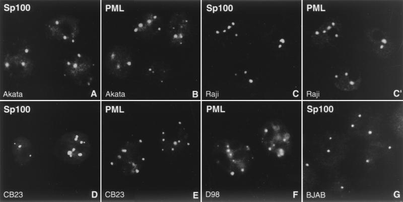
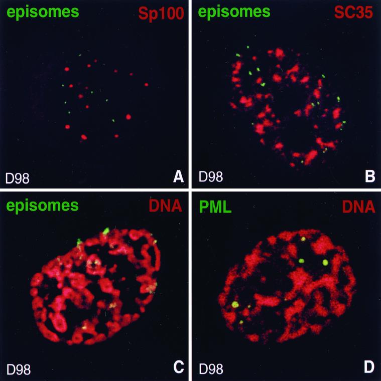
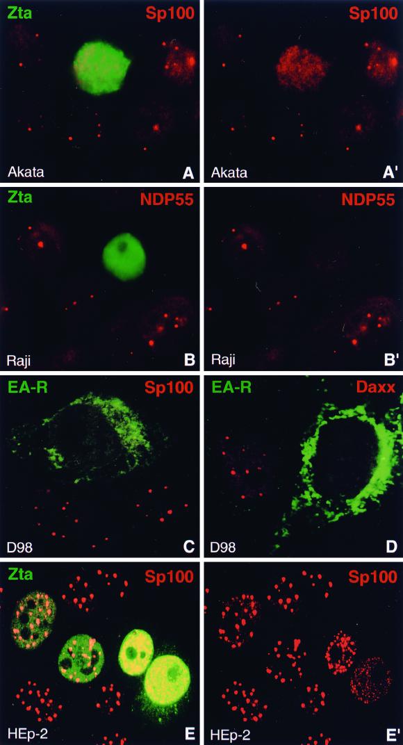
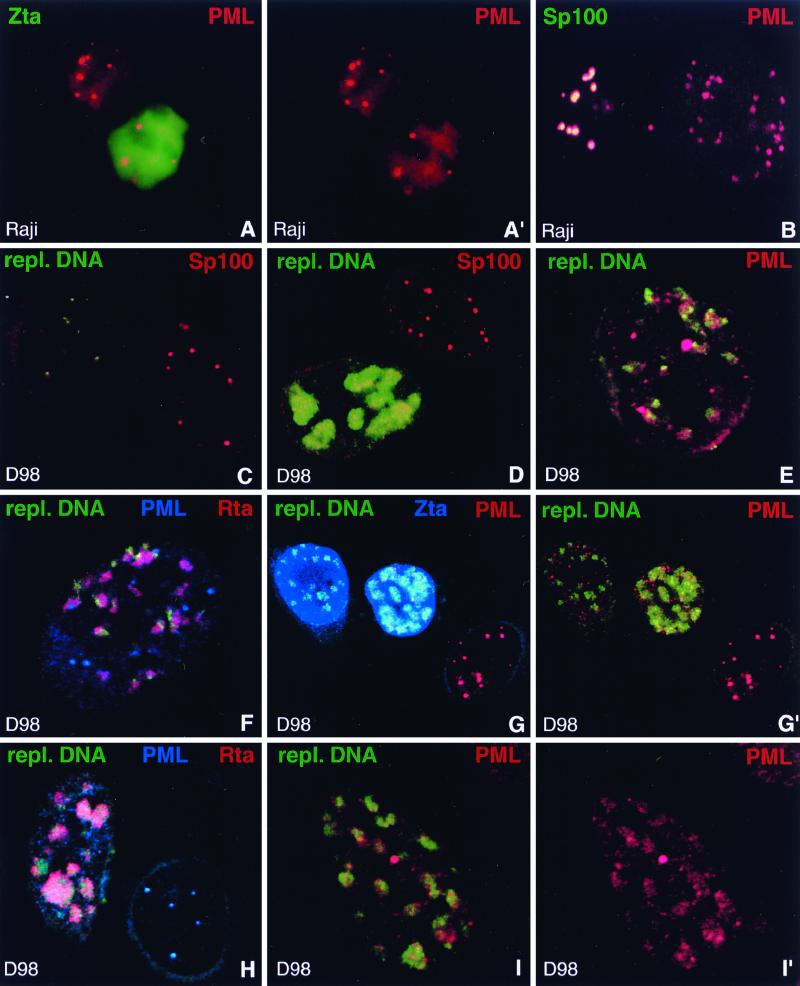
Nuclear domains called ND10 (nuclear domain 10) are discrete nuclear protein aggregations characterized by a set of interferon-upregulated proteins including Sp100 and PML, where papova-, adeno-, and herpesviruses begin their transcription and DNA replication. Both the alpha- and betaherpesvirus subfamilies disrupt ND10 upon infection by dispersing and/or destroying ND10-associated proteins. We studied the effect of the gammaherpesvirus Epstein-Barr virus (EBV) on ND10 and its spatial distribution in the nucleus of cells during latency and lytic reactivation. In latently infected Burkitt's lymphoma, lymphoblastoid, and D98/HR1 cells, ND10 were intact, as judged by immunofluorescence localization of PML, Sp100, NDP55, and Daxx. Fluorescent in situ hybridization revealed no association between viral episomes and ND10 during latency, implying that the maintenance replication of EBV, which depends on host cell proliferation, occurs independent of ND10. As in mitosis, the EBV genomes were attached to interphase chromosomes, suggesting that they are unable to move freely within the interchromosomal space and thus unable to associate with the interchromosomally located ND10 or other nuclear domains. Upon lytic activation, ND10 became dispersed in cells expressing lytic proteins. Redistribution of ND10 proteins occurred sequentially at different stages of the lytic cycle, with Sp100, Daxx, and NDP55 dispersed before and PML dispersed after the onset of lytic replication. ND10 remnants were retained until the early stages of lytic replication, and replicating EBV genomes were frequently found beside this nuclear domain; the number of replication domains was usually lower than the average latent virus frequency. Thus, latency does not require or induce interaction of EBV with ND10 for transcription and replication, whereas lytic replication triggers dispersion of ND10 proteins and occurs in close association with PML aggregates. The required movement of chromosome-attached latent EBV episomes to ND10 after reactivation from latency might include physical release of the chromosome-bound episomes. Only episomes contacting ND10 after such a release might be able to begin lytic replication.


Figures

FIG. 1
FIG. 1
ND10 are not disrupted during latency. The EBV-positive Burkitt's lymphoma cell lines Akata (A and B) and Raji (C and C′), the latently EBV-infected lymphoblastoid cell line CB23 (D and E), and the EBV-positive fusion cell line D98/HR1 (D98; F) possess intact ND10, as revealed by immunohistochemistry with antibodies against Sp100 (A, C, D, and G) and PML (B, C′, E, and F). C and C′ shows double labeling to demonstrate colocalization of both proteins. As a control, ND10 are visualized in the EBV-negative Burkitt's lymphoma cell line BJAB by staining for Sp100 (G).
FIG. 2
FIG. 2
Localization of EBV genomes during latency. The upper corner of each image shows the color used for labeling of the respective protein or DNA. (A) EBV episomes are not associated with ND10 during latency. ND10 are shown in a latently infected D98/HR1 cell by labeling Sp100, and episomes were visualized by FISH. (B) D98/HR1 cell during EBV latency stained with anti-SC35 and hybridized with an EBV-specific probe. Episomes are excluded from and only occasionally associated with SC35 domains. (C) Latently infected D98/HR1 cell treated to undergo SICC during interphase. Cellular DNA was stained with propidium iodide to visualize the contracted chromosomes, and in situ hybridization was performed with an EBV-specific probe for the detection of episomes. Episomes are found exclusively closely attached to the condensed interphase chromosomes. (D) As for panel C, but stained for PML to visualize ND10. In contrast to EBV episomes, ND10 are located between the chromosomes and thus occupy a different nuclear space.
FIG. 3
FIG. 3
Lytic activation of EBV disperses the ND10 proteins Sp100, NDP55, and Daxx. (A and A′) Lytic activation of EBV was induced in Akata cells and monitored by staining for the immediate-early protein Zta. Sp100 is no longer concentrated in ND10 in Zta-expressing cells. A′, Sp100 staining only. (B and B′) Lytic activation of EBV in Raji cells, detected by staining of Zta, induces the dispersion of the ND10 protein NDP55. B′, NDP55 staining only. (C) D98/HR1 cells were Zta transfected to induce lytic activation of EBV and stained for the viral protein EA-R and Sp100. EA-R, which is localized in the cytoplasm, allows the identification of cells in which EBV has entered the lytic cycle. Sp100 is no longer concentrated in ND10 after lytic activation. (D) Like Sp100 and NDP55, the ND10 protein Daxx disperses in EA-R-positive D98/HR1 cells. (E and E′) EBV-free HEp-2 cells were transfected with a Zta-expressing plasmid and stained with antibodies against Zta and Sp100. ND10 remain intact at Zta concentrations comparable to those observed after lytic activation, but Sp100 starts to disperse when Zta is expressed at very high levels (cells on right). Note the deposition of Zta in the cytoplasm in the lower right cell. E′, Sp100 staining only.
FIG. 4
FIG. 4
Behavior of PML upon EBV reactivation compared to Sp100. (A and A′) Raji cells stained for Zta and PML after induction of lytic activation. Unlike other ND10 proteins, PML often remains concentrated in ND10 in Zta-positive cells. A′, PML staining only. (B) Raji cells double labeled with antibodies against PML and Sp100 after disruption of latency to demonstrate release of Sp100 but not of PML from ND10. The left-hand cell shows typical ND10 which contain both proteins, indicating that EBV remained latent. In the right-hand cell, EBV apparently entered the lytic cycle and released Sp100 from ND10 which still contain PML. (C) D98/HR1 cells were Zta transfected to induce lytic activation, stained for Sp100, and probed with a labeled PCR product which recognizes clusters of replicating EBV DNA but not individual episomes. Note the lack of detectable hybridization signal in the right-hand cell, in which no lytic activation occurred. This cell contains Sp100-positive ND10. In contrast, the left-hand cell displays small, i.e., early, foci of replicating EBV DNA. Sp100 is no longer concentrated in ND10 as soon as replicated EBV genomes are detectable. (D) Zta-transfected D98/HR1 cells labeled for Sp100 and hybridized with the probe specific for replicating EBV DNA. The lower cell shows an advanced stage of the lytic cycle, with replicating EBV genomes that form large clusters of DNA. Sp100 remains dispersed and does not associate with the replication domains. In the upper cell, no lytic cycle has been induced and Sp100 is not dispersed from ND10. No replicated EBV DNA is detectable in this cell. (E) A Zta-transfected D98/HR1 cell was stained for PML and probed for replicating EBV DNA. In contrast to Sp100, PML-positive ND10 are still visible after lytic replication has started. Most of the replication domains are found in close association with PML. (F) D98/HR1 cells were transfected with Zta to induce lytic activation and triple labeled for PML and Rta by immunostaining and for replicating EBV genomes by FISH. Rta forms aggregates in close association with the replication domains but without complete colocalization. PML is still visible in the form of several ND10-like structures. (G and G′) Zta-transfected D98/HR1 cells triple labeled for the detection of PML, Zta, and replicating EBV DNA. The replication-specific probe detects EBV genomes only in Zta-positive cells (two leftmost cells). In the right-hand untransfected cell, PML is almost completely aggregated into ND10, whereas PML becomes increasingly dispersed during lytic replication (two leftmost cells). However, even during late replication stages, as represented by the middle cell, PML is still concentrated in several ND10-like foci. In panel G′, Zta staining is omitted. (H) Zta-transfected D98/HR1 cell immunostained for Rta and PML and probed for replicating EBV DNA. In the right-hand cell, which is probably not transfected, no lytic activation was induced. The replication-specific probe fails to recognize EBV-specific genomes in this cell, which displays intact PML-positive ND10. The left-hand cell represents a late stage of the lytic cycle, as judged from the size of the replication compartments. PML foci have almost completely disappeared. In contrast to earlier replication stages as shown in panel F, the Rta aggregations and replication domains show a higher degree of colocalization, suggesting that both components had merged while increasing in size. (I and I′) Zta-transfected D98/HR1 cell hybridized with the replication-specific probe and immunostained for PML. PML is redistributed into the replication domains and colocalizes almost completely with the replicated EBV genomes in the form of diffuse PML accumulations. I′, PML staining only.

References

    1. Adamson A L, Kenney S. The Epstein-Barr virus BZLF1 protein interacts physically and functionally with the histone acetylase CREB-binding protein. J Virol. 1999;73:6551–6558. - PMC - PubMed
    1. Ahn J H, Hayward G S. The major immediate-early proteins IE1 and IE2 of human cytomegalovirus colocalize with and disrupt PML-associated nuclear bodies at very early times in infected permissive cells. J Virol. 1997;71:4599–4613. - PMC - PubMed
    1. Ahn J H, Jang W J, Hayward G S. The human cytomegalovirus IE2 and UL112–113 proteins accumulate in viral DNA replication compartments that initiate from the periphery of promyelocytic leukemia protein-associated nuclear bodies (PODs or ND10) J Virol. 1999;73:10458–10471. - PMC - PubMed
    1. Ascoli C A, Maul G G. Identification of a novel nuclear domain. J Cell Biol. 1991;112:785–795. - PMC - PubMed
    1. Baer R, Bankier A T, Biggin M D, Deininger P L, Farrell P J, Gibson T J, Hatfull G, Hudson G S, Satchwell S C, Seguin C, et al. DNA sequence and expression of the B95–8 Epstein-Barr virus genome. Nature. 1984;310:207–211. - PubMed

Publication types