. 2000 Nov;12(11):2047-60.
doi: 10.1105/tpc.12.11.2047.

Cell wall alterations in the arabidopsis emb30 mutant

Affiliations

Cell wall alterations in the arabidopsis emb30 mutant

D E Shevell et al. Plant Cell. 2000 Nov.

Abstract

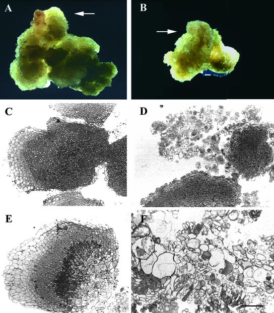
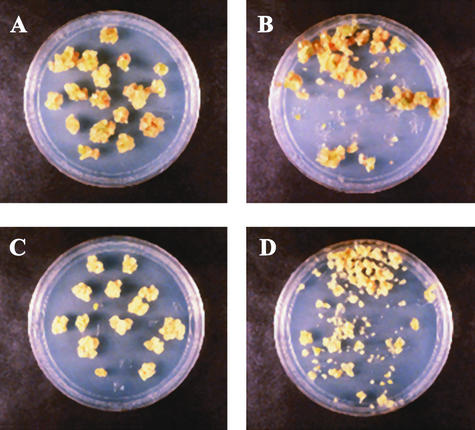
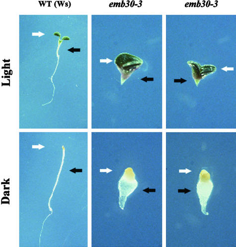
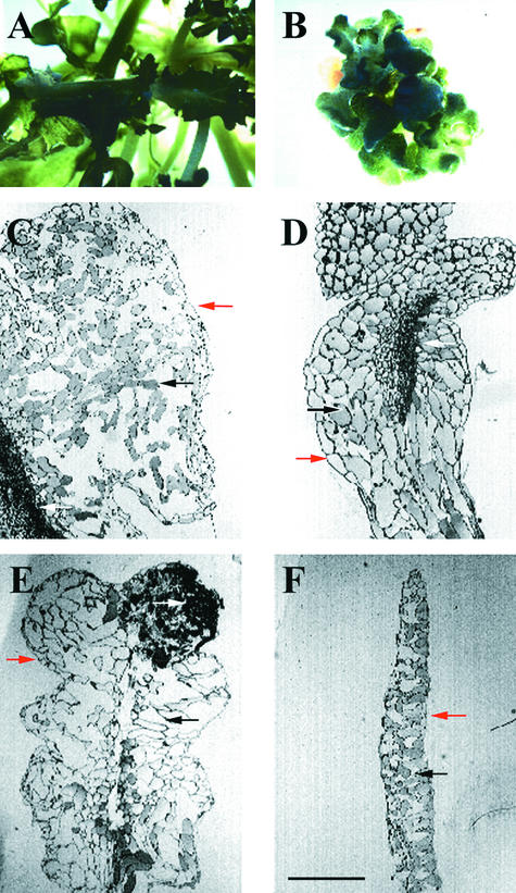
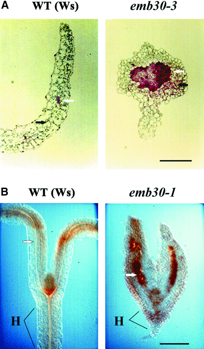
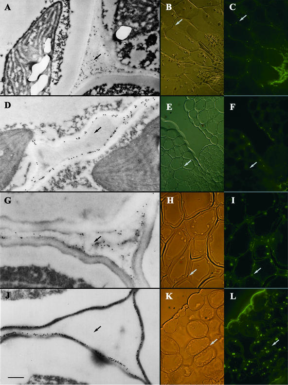
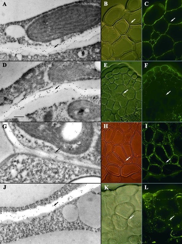
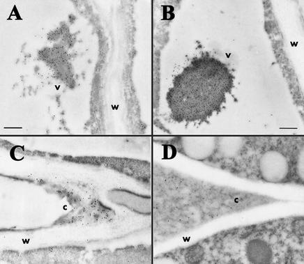
The Arabidopsis EMB30 gene is essential for controlling the polarity of cell growth and for normal cell adhesion during seedling development. In this article, we show that emb30 mutations also affect the growth of undifferentiated plant cells and adult tissues. EMB30 possesses a Sec7 domain and, based on similarities to other proteins, presumably functions in the secretory pathway. The plant cell wall depends on the secretory pathway to deliver its complex polysaccharides. We show that emb30 mutants have a cell wall defect that sometimes allows material to be deposited into the interstitial space between cells instead of being restricted to cell corners. In addition, pectin, a complex polysaccharide important for cell adhesion, appears to be abnormally localized in emb30 plants. In contrast, localization of epitopes associated with xyloglucan or arabinogalactan was similar in wild-type and emb30 tissues, and the localization of a marker molecule to vacuoles appeared normal. Therefore, emb30 mutations do not cause a general defect in the secretory pathway. Together, these results suggest that emb30 mutations result in an abnormal cell wall, which in turn may account for the defects in cell adhesion and polar cell growth control observed in the mutants.


Figures

Figure 1.
Figure 1.
EMB30 Is Required for Normal Growth of Callus. Seeds were germinated on MS medium (see Methods); 14-day-old seedlings were transferred to CIM and harvested 29 days later. Results were similar for seedlings germinated directly on CIM (data not shown). (A) and (B) Wild-type and emb30-3 calluses, respectively. Arrows point to one of several areas of callus growth. (C) Wild-type Wassilewskija (Ws) callus. (D) emb30-4 callus. (E) Wild-type Ws callus. (F) emb30-3 callus. The emb30 sections shown have weak (D) or severe (F) phenotypes representative of all the emb30 alleles examined (emb30-1, emb30-3, and emb30-4). (A) and (B) are at the same magnification (×4); (C) to (F) are at the same magnification. formula image.
Figure 2.
Figure 2.
emb30 Mutant Calluses Are More Friable Than Those of Wild Type. Calluses were subjected to agitation as described in Methods. (A) and (B) Wild-type Ws before and after treatment, respectively. (C) and (D) emb30-3 before and after treatment, respectively.
Figure 3.
Figure 3.
Response of emb30 Mutants to Light or Darkness. Wild-type and emb30-3 seedlings grown in 16-hr-light/8-hr-dark periods or in the dark for 7 days. The light-grown seedlings are green; the dark-grown seedlings are white-yellow. Magnification of wild-type Ws seedlings is ×2; that of emb30-3 seedlings is ×4. The white arrows point to the cotyledons; the black arrows point to the hypocotyls. Similar results were obtained with emb30-1 (data not shown).
Figure 4.
Figure 4.
Wild-Type and emb30 Leaves on SIM. Phenotypes similar to those shown were observed in all emb30 alleles examined (emb30-1, emb30-2, emb30-3, and emb30-4). Red arrows point to epidermal cells; black arrows point to mesophyll cells; white arrows point to vascular-like cells. (A) and (B) Wild-type and emb30-1 leaves, respectively, grown on SIM. (C) emb30-2 leaf. (D) emb30-1 leaf. (E) emb30-4 leaf. (F) Wild-type leaf. (A) and (B) are at the same magnification (×3); (C) to (F) are at the same magnification. formula image.
Figure 5.
Figure 5.
Histochemical Localization of Pectin in Wild-Type and emb30 Seedlings. (A) RR-stained sections. (B) HF-AC–stained sections of tissue pretreated with hot acidic methanol (see Methods). Black arrows point to mesophyll cells; white arrows point to vascular cells. emb30-1 and emb30-3 seedlings were each examined with both types of stains (this figure and data not shown). H, hypocotyl; WT, wild type. formula image; bformula image for both wild-type and emb30 seedlings.
Figure 6.
Figure 6.
Immunostaining of emb30-3 and Wild-Type Cotyledon Sections with JIM5 and PGA/RG-I Antibodies. (A) to (C) JIM5 staining of emb30-3 section. JIM5 stains the cell wall and often, in emb30-3 mutants, the interstitial space between cells. (D) to (F) JIM5 staining of wild-type sections. (G) to (I) PGA/RG-I antibody staining of emb30-3 sections. PGA/RG-I antibody also stains the cell wall and, in emb30-3 sections, the interstitial space between cells. (J) to (L) PGA/RG-I antibody staining of wild-type sections. (A), (D), (G), and (J) show IEM of the sections. (B), (E), (H), and (K) show Nomarski optics and (C), (F), (I), and (L) show IF microscopy of the same sections, respectively. Arrows point to the interstitial space between cells. formula image.
Figure 7.
Figure 7.
Immunostaining of emb30-3 and Wild-Type Cotyledon Sections with CCRC-M1 and CCRC-M7 Antibodies. (A) to (C) CCRC-M1 staining of emb30-3 sections. (D) to (F) CCRC-M1 staining of wild-type sections. emb30-3 and wild-type sections show a similar pattern of staining with CCRC-M1. (G) to (I) CCRC-M7 staining of emb30-3 sections. (J) to (L) CCRC-M7 staining of wild-type sections. emb30-3 and wild-type sections show a similar pattern of staining with CCRC-M7. (A), (D), (G), and (J) show IEM of sections. Arrows point to the cell wall. (B), (E), (H), and (K) show Nomarski optics and (C), (F), (I), and (L) show IF microscopy of the same sections, respectively. In (B), (C), (E), (F), (H), (I), (K), and (L), arrows point to the interstitial space between cells. formula image.
Figure 8.
Figure 8.
IEM with an Antibody That Recognizes Lectin. (A) Wild-type Ws seedling expressing CTPP+ lectin. (B) emb30-3 seedling expressing CTPP+ lectin. (C) Wild-type seedling expressing CTPP− lectin. (D) emb30-3 seedling expressing CTPP− lectin. c, cell corner; v, vacuole; w, cell wall. (A), (C), and (D) are at the same magnification. formula image; formula image.

References

    1. Altschul, S.F., Madden, T.L., Schaffer, A.A., Zhang, J., Zhang, Z., Miller, W., and Lipman, D.J. (1997). Gapped BLAST and PSI-BLAST: A new generation of protein database search programs. Nucleic Acids Res. 25, 3389–3402. - PMC - PubMed
    1. Baus, A.D., Franzmann, L., and Meinke, D.W. (1986). Growth in vitro of arrested embryos from lethal mutants of Arabidopsis thaliana. Theor. Appl. Genet. 72, 577–586. - PubMed
    1. Belanger, K.D., and Quatrano, R.S. (2000). Polarity: The role of localized secretion. Curr. Opin. Plant Biol. 3, 67–72. - PubMed
    1. Bouget, F., Berger, F., and Brownlee, C. (1998). Position dependent control of cell fate in the Fucus embryo: Role of intercellular communication. Development 125, 1999–2008. - PubMed
    1. Busch, M., Mayer, U., and Jürgens, G. (1996). Molecular analysis of the Arabidopsis pattern formation gene GNOM: Gene structure and intragenic complementation. Mol. Gen. Genet. 250, 681–691. - PubMed

Publication types