. 2000 Nov;12(11):2075-86.
doi: 10.1105/tpc.12.11.2075.

Oriented asymmetric divisions that generate the stomatal spacing pattern in arabidopsis are disrupted by the too many mouths mutation

Affiliations

Oriented asymmetric divisions that generate the stomatal spacing pattern in arabidopsis are disrupted by the too many mouths mutation

M Geisler et al. Plant Cell. 2000 Nov.

Abstract

Wild-type stomata are spaced by intervening cells, a pattern disrupted in the Arabidopsis mutant too many mouths (tmm). To determine the mechanism of wild-type spacing and how tmm results in pattern violations, we analyzed the behavior of cells through time by using sequential dental resin impressions. Meristemoids are stomatal precursors produced by asymmetric division. We show that wild-type patterning largely results when divisions next to a preexisting stoma or precursor are oriented so that the new meristemoid is placed away. Because this placement is independent of cell lineage, these divisions may be oriented by cell-cell signaling. tmm randomizes this orientation and releases a prohibition on asymmetric division in cells at specific locations, resulting in stomatal clusters. TMM is thus necessary for two position-dependent events in leaves: the orientation of asymmetric divisions that pattern stomata, and the control of which cells will enter the stomatal pathway. In addition, our findings argue against most previous hypotheses of wild-type stomatal patterning.


Figures

Figure 1.
Figure 1.
Overview of Cell Types. The stomatal lineage begins when a meristemoid mother cell (MMC; gray shading) forms and then divides asymmetrically, producing a meristemoid and a larger sister cell. Meristemoids divide asymmetrically zero to three times and then convert into guard mother cells (GMCs). The latter divide symmetrically, producing two guard cells. Smaller, less sinuous epidermal cells (upper left) become MMCs and divide asymmetrically (i), divide symmetrically (ii), or form wavy pavement cells (iii). Cells adjacent to a stoma or precursor can initiate a new stomatal lineage (iv and v), as can cells that are not adjacent. The first division of an MMC located next to a stoma or precursor is oriented so that the resulting satellite meristemoid is placed away (bottom left). Satellite meristemoids can divide zero to three times (only one division is illustrated in the bottom row). Neighbor cells that are clonally related to the stoma (see text) are drawn with solid rather than dashed lines (top row). The three stomatal complexes shown at top right are anisocytic. Div., division; M, meristemoid; SM, satellite meristemoid.
Figure 2.
Figure 2.
Key Events in Stomatal Development Shown in a Dental Impression Series. The abaxial epidermis of a single cotyledon is shown through time. Cells c (day 7) and f (day 8) are MMCs because they later divided asymmetrically to produce meristemoids. The division of MMC f took place next to a preexisting stoma and produced a satellite meristemoid. Both MMCs arose from smaller, less sinuous cells. One smaller cell (e; day 7) divided symmetrically by day 8. Meristemoid a (day 6) divided twice asymmetrically in an inward spiral. Two apparent meristemoids are adjacent (day 6, lower right); the upper one formed a stoma, but the lower meristemoid (b) did not progress in development (d). Blue, MMC; red, meristemoid; yellow, GMC; green, stoma.
Figure 3.
Figure 3.
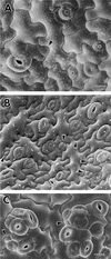
Cryoscanning Electron Microscopy of Wild-Type Patterning and the Disruption of Stomatal Spacing in tmm. (A) Wild type. Both the satellite meristemoid (arrowhead) and the larger sister cell (at left) were derived from an asymmetric division of an MMC that was adjacent to a stoma or precursor (now a mature stoma with a pore). Two GMCs (asterisks) were each derived from a satellite meristemoid. Successive stages of pore development are shown at lower and upper right. Note that five of the six existing and future stomata shown are patterned by satellite meristemoid placement. (B) Wild type. Arrowheads indicate satellite meristemoids, some of which have divided. Arrow shows nonsatellite meristemoid or GMC. (C) Stomatal clusters in tmm. The arrowheads indicate incorrectly placed satellite meristemoids. Many of the stomata in the clusters are in various stages of pore formation. formula image; formula image.
Figure 4.
Figure 4.
Asymmetric Divisions: Unequal in Both Cell Size and Fate. The ratio of daughter cell size in divisions with an asymmetric fate (MMCs; circles) and with a symmetric fate (divisions of smaller cells that produced two pavement cells; squares). No divisions that were asymmetric in fate had daughter cells of equal size, and no divisions that were symmetric in fate had daughter cells differing in size by >30%. Measurements of daughter cell size were made from dental resin sequences of 300 divisions that were known to be asymmetric in fate and of 100 that were symmetric in fate. Curves represent best fit.
Figure 5.
Figure 5.
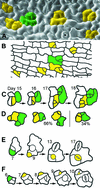
Placement and Divisions of MMCs. (A) Scanning electron microscopy of a dental impression from a wild-type cotyledon (4 days after germination), showing that some MMCs are not spaced apart. Each group of cells delimited by color fill came from a single MMC, as deduced from other replicas in the series. (B) As in (A) except that MMC lineages were deduced from cell wall positions. Shown is a tracing from a fixed cotyledon (12 hr after germination). (C) Divisions in adjacent MMCs are randomly placed. A dental impression sequence shows divisions of adjacent MMCs that resulted in nonadjacent meristemoids (day 16). Green and yellow areas delimit lineages from each MMC. (D) As in (C). Sixty-six percent of the divisions of adjacent MMCs yielded separated meristemoids but 34% produced meristemoids in contact. Drawings, which represent different positions, are each from a different dental resin series and summarize results from a sample of fifty adjacent MMCs. (E) Dental resin series showing that satellite meristemoids are placed away from previously formed precursor cells. The asymmetric division of the MMC (green cell) is oriented so that the new meristemoid (yellow cell at day 9) is separated from the preexisting precursor cell (a meristemoid at day 7; a GMC at day 9) by an intervening sister cell. (F) As in (E). Both meristemoids divide asymmetrically. formula image.
Figure 6.
Figure 6.
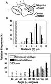
Wild-Type Satellite Meristemoids Are Correctly Placed Regardless of Cell Lineage, and Orientation Is Randomized in tmm. (A) Measurement of angle (α) and initial distance (Δ) of satellite meristemoid from preexisting stoma, GMC, or meristemoid (see Methods). (B) Almost all wild-type satellite meristemoids were placed at least 4 μm away regardless of whether the MMC that produced them was clonally related to the preexisting stoma or precursor. Many tmm satellite meristemoids were closer than 4 μm. (C) Distribution of angles of tmm but not wild-type satellite meristemoids is random.
Figure 7.
Figure 7.
Dental Resin Sequence Showing Correct Placement of Satellite Meristemoid from Nonclonal MMC. Shown at left is a young cotyledon (144 hr after germination) with cell files still detectable (stippling). File and cell lineage of MMC (+) differ from those of the adjacent meristemoid (left of MMC). Division results in correct placement of the satellite meristemoid (asterisk). At right, asymmetric division of the satellite meristemoid is shown. Bar = 50 μm.
Figure 8.
Figure 8.
Responses of Adjacent Meristemoids That Create One-Celled Spacing. (A) On one occasion, both meristemoids were observed to convert into GMCs, producing two stomata that are in contact. (B) Left meristemoid divides away. (C) Right meristemoid differentiates into a pavement cell. The lightly and darkly stippled cells denote different meristemoids and their respective derivatives.
Figure 9.
Figure 9.
Position-Dependent Control of Cell Fate and Disruption by tmm. Relative frequencies with which smaller, less sinuous cells followed three different fates as a function of the number of adjacent stomata, GMCs, or meristemoids (0 to 3, singly or in combination). Wild-type (WT) cells adjacent to two stomata or precursors did not divide; they usually differentiated into pavement cells. Most tmm cells adjacent to one or more stomata or precursors divided asymmetrically regardless of the number of adjacent cells. NA, not applicable.
Figure 10.
Figure 10.
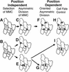
Overview of Position-Dependent and Position-Independent Events. (A) and (B) The placement of the first precursor cell, the MMC, is random. (C) The placement of asymmetric divisions of separated MMCs appears to be random. (D) and (E) Divisions of MMCs in contact are randomly oriented and can produce meristemoids in contact. (F) The orientation of asymmetric divisions in neighbor cells depends upon the position of the stoma or precursor. (G) Asymmetric divisions of adjacent meristemoids can be oriented and thus space stomata. (H) Cells next to two stomata or precursors (asterisks) follow a pavement cell fate, but this does not directly create the one-celled spacing. (I) Adjacent meristemoids can become pavement cells (asterisk) and thus space stomata. MMCs are shown as stippled regions in (A) and (B). Meristemoids are dark gray.
Figure 11.
Figure 11.
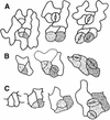
Mechanisms of tmm Stomatal Cluster Formation Shown in Dental Resin Series. (A) Six of the seven satellite meristemoids shown (stippled) are misplaced and contact preexisting stomata or precursor cells. (B) Some cells between two stomata or precursors (arrowheads) divide asymmetrically. (C) A sister cell to a satellite meristemoid becomes a GMC (asterisks) without any division. (D) Both adjacent meristemoids (stippled) develop into stomata.

Similar articles

Cited by

References

    1. Berger, D., and Altmann, T. (2000). A subtilisin-like serine protease involved in the regulation of stomatal density and distribution in Arabidopsis thaliana. Genes Dev. 14, 1119–1131. - PMC - PubMed
    1. Bünning, E. (1956). General processes of differentiation. In The Growth of Leaves, F.L. Milthorpe, ed (London: Butterworths Scientific Publications), pp. 18–30.
    1. Croxdale, J.L. (2000). Stomatal patterning in angiosperms. Am. J. Bot. 87, 1069–1080. - PubMed
    1. Drubin, D.G., and Nelson, W.J.. (1996). Origins of cell polarity. Cell 84, 335–344. - PubMed
    1. Fowler, J.E., and Quatrano, R.S. (1997). Plant cell morphogenesis: Plasma membrane interactions with the cytoskeleton and cell wall. Annu. Rev. Cell Dev. Biol. 13, 697–743. - PubMed

Publication types