. 2000 Nov;12(11):2129-42.
doi: 10.1105/tpc.12.11.2129.

Tobacco VDL gene encodes a plastid DEAD box RNA helicase and is involved in chloroplast differentiation and plant morphogenesis

Affiliations

Tobacco VDL gene encodes a plastid DEAD box RNA helicase and is involved in chloroplast differentiation and plant morphogenesis

Y Wang et al. Plant Cell. 2000 Nov.

Abstract

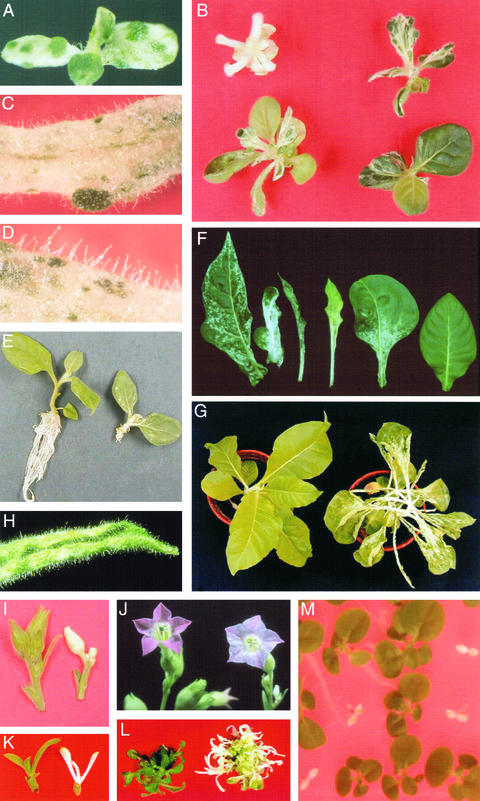
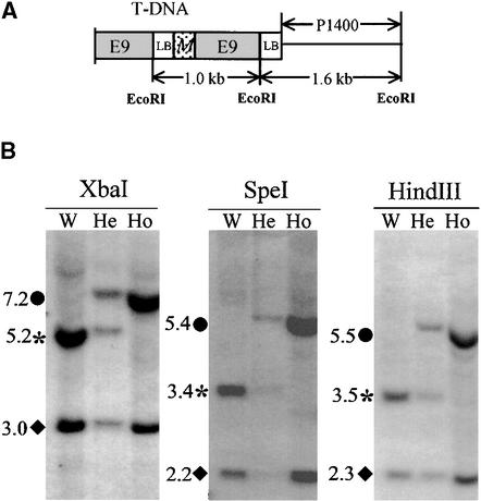
The recessive nuclear vdl (for variegated and distorted leaf) mutant of tobacco was obtained by T-DNA insertion and characterized by variegated leaves and abnormal roots and flowers. Affected leaf tissues were white and distorted, lacked palisadic cells, and contained undifferentiated plastids. The variegation was due to phenotypic, rather than genetic, instability. Genomic and cDNA clones were obtained for both the mutant and wild-type VDL alleles. Three transcripts, resulting from alternate intron splicing or polyadenylation, were found for the wild type. The transcripts potentially encode a set of proteins (53, 19, and 15 kD) sharing the same N-terminal region that contains a chloroplast transit peptide capable of importing the green fluorescent protein into chloroplasts. The predicted 53-kD product belongs to the DEAD box RNA helicase family. In the homozygous vdl mutant, T-DNA insertion resulted in accumulation of the shortest transcript and the absence of the RNA helicase-encoding transcript. Genetic transformation of the homozygous mutant by the 53-kD product-encoding cDNA fully restored the wild-type phenotype. These data suggest that a plastid RNA helicase controls early plastid differentiation and plant morphogenesis.


Figures

Figure 1.
Figure 1.
Phenotype of the vdl Mutant. (A) A vdl seedling grown in vitro in Murashige and Skoog (MS) agar medium (see Methods). (B) Four vdl plants grown in vitro and showing various phenotypes. The seedlings (5 weeks old) were grown in MS agar medium. (C) and (D) Details of a vdl plant grown in vitro. (E) Wild-type (left) and vdl (right) plants grown in vitro (4 weeks old). (F) Leaves from various vdl plants grown in soil, showing various phenotypes. (G) Wild-type (1 month old, at left) and vdl (2 months old, at right) plants grown in soil. (H) Detail of a narrow leaf of a vdl plant similar to that shown in (G). (I) Flower bud from wild-type (left) and vdl (right) plants. (J) Flower from wild-type (left) and vdl (right) plants. (K) Development of an axillary bud on green stem sections derived from wild-type (left) and vdl (right) plants and transferred for 2 weeks to MS agar medium. (L) Leaf discs from wild-type (left) and vdl (right) green leaves transferred to an MS agar medium for shoot regeneration. (M) A transgenic line (T6-5-8) resulting from the transformation of a vdl homozygous plant with a wild-type VDL-1 cDNA clone was grown until the flowering stage and then self-pollinated; seed was then sown on a growth medium without antibiotics. The photograph was taken 5 weeks after sowing.
Figure 2.
Figure 2.
Light Microscopy Analysis of Wild-Type and Homozygous vdl Leaves and Roots. (A) Cross-section from a green sector of a vdl leaf. (B) Cross-section from a white sector of a vdl leaf. (C) Cross-section from a curled vdl leaf similar to that shown in Figure 1H. (D) Cross-section from a vdl leaf at a junction between a green (left) or white (right) sector. (E) Cross-section from a white spot within a green sector of a vdl leaf. (F) Cross-section from a vdl root. The epidermis and the endodermis are not well identified, and the vascular cylinder has a disorganized structure. (G) Cross-section from a wild-type root. formula image.
Figure 3.
Figure 3.
Transmission Electron Micrographs of vdl Leaf Cells. (A) Green sector of a vdl leaf. The plastid structure is similar to that found for the wild type shown in (C). (B) White sector of a vdl leaf. Plastids show a vesicular instead of lamellar structure. (C) Wild-type leaf. Chloroplasts show the typical stacking thylakoids. formula image (A) formula image (A) formula image (C).
Figure 4.
Figure 4.
A T-DNA−Flanking Genomic Fragment Cosegregates with the vdl Mutation. (A) Structure of the cosegregating T-DNA insert. The recombined T-DNA insert consisted of two copies of the T-DNA left border (LB), part of the MIP gene (M), and the E9 terminator. The cloned 1.6- and 1.0-kb EcoRI T-DNA fragments and the 1.4-kb genomic fragment (P1400) are indicated. (B) Genomic DNA was extracted from wild-type (W), heterozygous (He), and homozygous (Ho) vdl plants and digested with the indicated restriction enzymes. After transfer, the DNA was hybridized with the primer-generated probe P1400. Circles indicate the vdl locus, asterisks indicate the wild-type VDL locus, and diamonds indicate the VDL′ locus.
Figure 5.
Figure 5.
Genomic and cDNA Organization of the VDL and VDL′ Loci. (A) Genomic map of the VDL and VDL′ loci showing exons (numbered black boxes), introns (open boxes), and the location of the T-DNA insert. EcoRI (E), HindIII (H), and XbaI (X) restriction sites are indicated. Because of the considerable difference in the 5′ untranslated region, the first exons of the VDL and VDL′ loci are named 1 and 1′. The hatched region in VDL′ represents an upstream sequence that is completely different from that in VDL. The size of VDL introns 3 and 9 is based on restriction mapping (some gaps in the sequence remain). The sequence downstream of VDL′ exon 10 has not been cloned. (B) Alternative splicing products of the VDL and VDL′ loci. The filled boxes indicate exons. The asterisks in front of exons 5, 6, and 11 in VDL-6, VDL-7, and VDL-8, indicate that the 5′ or 3′ borders differed from those in the other cDNA clones, resulting in shorter transcripts; the exact sequence can be found in the databases.
Figure 6.
Figure 6.
RT-PCR Transcript Analysis of the VDL and VDL′ Loci. (A) Poly(A)+ RNA from wild-type leaves was amplified by RT-PCR. The 5′ primers were located in the 5′ untranslated region and were specific to either VDL (pTA5*A) or VDL′ (pTABis5*A); the 3′ primers were common to VDL and VDL′ and were located in exon 11 (pTFL65B). (B) Poly(A)+ RNA from wild-type (W), heterozygous (He), and homozygous (Ho) (white sections) mutant leaves was amplified by RT-PCR. The 5′ primers were as in (A); the 3′ primers were common to VDL and VDL′ and corresponded to exon 11 (pTFL57), exon 2b (pRH14), or exon 3 (pRH11). (C) Poly(A)+ RNA from wild-type flower (F), leaf (L), or root (R) was amplified by nested RT-PCR with, for VDL, primers pTA5A and pTFL50 (5′) and pTFL57 (3′), and for VDL′, primers pTABis5A (5′) and pTFL65B and pTFL57 (3′). Note that, to simplify the pattern, the 3′ primer used here for exon 11 was chosen to cover a region not present in VDL′-6, VDL′-7, and VDL′-8, which have a shorter exon 11 because of their alternative splicing. The numbers at left and right denote molecular mass markers (kilobases).
Figure 7.
Figure 7.
Alignment of the VDL-1/VDL′ -1 Protein Sequence with Sequences of Similar RNA Helicases. Sequence alignment showing the conserved blocks and the consensus sequence of the aligned proteins. The seven motifs (I, Ia, II, III, IV, V, and VI) of DEAD box RNA helicases are indicated by asterisks. A conserved region of unknown function between motifs Ia and II is also indicated. Boldface letters indicate amino acids that are 100% conserved. Amino acid residues of the consensus sequence are in lowercase when conserved in at least 70% of the proteins displayed or in uppercase when they are 100% conserved. The number symbol (#) indicates the hydrophobic amino acid Met, Ile, Leu, or Val. aa, amino acids.
Figure 8.
Figure 8.
Predicted VDL Transit Peptide Imports the GFP into Chloroplasts. The sequence coding for the first 73 amino acid residues of VDL-1 (VDL73) was inserted between the CaMV 35S promoter and the GFP sequence to give 35S-VDL73-GFP in a bacterial plasmid (see Methods). A 35S-GFP construct without the VDL sequence was used as a control. Both plasmids were electroporated into tobacco leaf protoplasts, and the fluorescence was analyzed after 24 hr with a confocal microscope. (A) GFP fluorescence in a protoplast transformed with 35S-VDL73-GFP. (B) Chlorophyll fluorescence of the protoplast shown in (A). (C) Merging of (A) and (B). (D) GFP fluorescence in a protoplast transformed with the control 35S-GFP.

Similar articles

Cited by

References

    1. Amaratunga, M., and Lohman, T.M. (1993). Escherichia coli rep helicase unwinds DNA by an active mechanism. Biochemistry 32, 6815–6820. - PubMed
    1. Bird, L.E., Subramanya, H.S., and Wigley, D.B. (1998). Helicases: A unifying structural theme? Curr. Opin. Struct. Biol. 8, 14–18. - PubMed
    1. Boddeker, N., Stade, K., and Franceschi, F. (1997). Characterization of DbpA, an Escherichia coli DEAD box protein with ATP independent RNA unwinding activity. Nucleic Acids Res. 25, 537–547. - PMC - PubMed
    1. Cabrera y Poch, H.L., Peto, C.A., and Chory, J. (1993). A mutation in the Arabidopsis DET3 gene uncouples photoregulated leaf development from gene expression and chloroplast biogenesis. Plant J. 4, 671–682.
    1. Carol, P., Stevenson, D., Bisanz, C., Breitenbach, J., Sandmann, G., Mache, R., Coupland, G., and Kuntz, M. (1999). Mutations in the Arabidopsis gene IMMUTANS cause a variegated phenotype by inactivating a chloroplast terminal oxidase associated with phytoene desaturation. Plant Cell 11, 57–68. - PMC - PubMed

Publication types

MeSH terms

LinkOut - more resources