. 2000 Nov;12(11):2201-18.
doi: 10.1105/tpc.12.11.2201.

A rab1 GTPase is required for transport between the endoplasmic reticulum and golgi apparatus and for normal golgi movement in plants

Affiliations

A rab1 GTPase is required for transport between the endoplasmic reticulum and golgi apparatus and for normal golgi movement in plants

H Batoko et al. Plant Cell. 2000 Nov.

Abstract

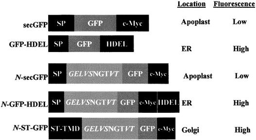
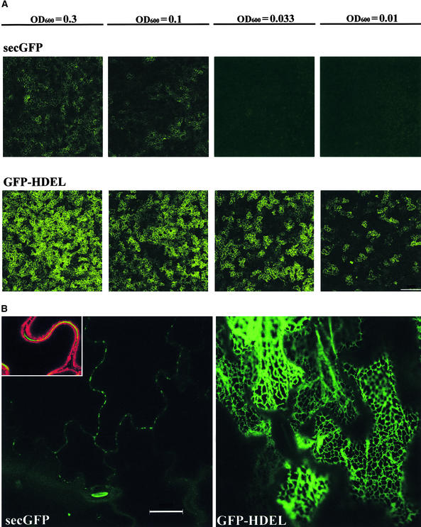
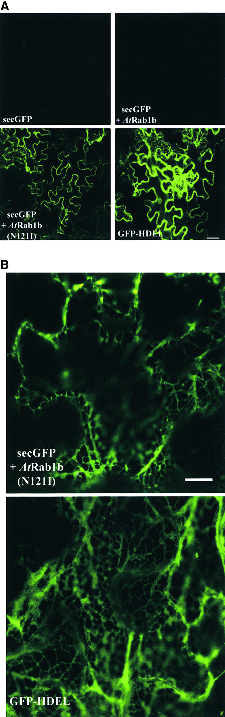
We describe a green fluorescent protein (GFP)-based assay for investigating membrane traffic on the secretory pathway in plants. Expression of AtRab1b(N121I), predicted to be a dominant inhibitory mutant of the Arabidopsis Rab GTPase AtRab1b, resulted in accumulation of a secreted GFP marker in an intracellular reticulate compartment reminiscent of the endoplasmic reticulum. This accumulation was alleviated by coexpressing wild-type AtRab1b but not AtRab8c. When a Golgi-targeted and N-glycosylated variant of GFP was coexpressed with AtRab1b(N121I), the variant also accumulated in a reticulate network and an endoglycosidase H-sensitive population appeared. Unexpectedly, expression of AtRab1b(N121I), but not of the wild-type AtRab1b, resulted in a reduction or cessation of vectorial Golgi movement, an effect that was reversed by coexpression of the wild type. We conclude that AtRab1b function is required for transport from the endoplasmic reticulum to the Golgi apparatus and suggest that this process may be coupled to the control of Golgi movement.


Figures

Figure 1.
Figure 1.
Constructs for Transient Expression. secGFP is derived from the ER-targeted GFP-HDEL by replacing the C-terminal tetrapeptide with a c-Myc epitope tag. N-secGFP, N-GFP- HDEL, and N-ST-GFP are derived from secGFP, GFP-HDEL, and ST-GFP (Boevink et al., 1998), respectively, by addition of an N-terminal peptide containing an N-glycosylation site. The steady state location and the fluorescence characteristics of each GFP in this location are indicated at right. All constructs were inserted under control of an enhanced CaMV promoter in an Agrobacterium binary vector (Hawes et al., 2000). NGT, single-letter amino acid code; ST, sialyltransferase.
Figure 2.
Figure 2.
Agrobacterium-Mediated Transient Expression of secGFP and GFP-HDEL. (A) Differential fluorescence signals from tobacco epidermal cells after transient expression of secGFP and GFP-HDEL using Agrobacterium strains infiltrated at various OD600 values, as indicated above each pair of images. Individual epidermal cells exhibiting GFP fluorescence in the apoplast (secGFP) and cortical cytoplasm (GFP-HDEL) can be seen. (Because the epidermal cells possess a large central vacuole with a thin layer of cortical cytoplasm, only the GFP-HDEL signal originating from the ER in the cortical cytoplasm adjacent to the anticlinal cell walls is clearly visible at this magnification.) The images are a projection of a 30 × 1-μm Z-series collected by confocal laser scanning microscopy. (B) High-resolution image of epidermal cells inoculated with secGFP and GFP-HDEL at OD600 0.01, collected by confocal laser scanning microscopy. The inset shows secGFP fluorescence in the apoplast (green) imaged in the presence of 5 μg/mL plasma membrane–labeling dye FM4-64 (red). To show the typical reticulate organization of the ER, the GFP-HDEL image is taken from an optical section passing through the cortical cytoplasm that lies immediately adjacent to the outer periclinal cell walls; the secGFP image is taken from a section passing through the center of the cells and shows the anticlinal walls in cross-section with their associated secGFP fluorescence. formula image; formula image.
Figure 3.
Figure 3.
secGFP Reports Reversible Inhibition of Membrane Traffic by BFA. (A) Confocal microscopy of GFP fluorescence in epidermal cells of samples of leaves infiltrated with Agrobacterium strains carrying either secGFP (left) or GFP-HDEL (right) after either 15 hr of incubation in water (Water) or 8 hr of incubation in 10 μg/mL BFA followed by washing and a further 7-hr incubation in either 10 μg/mL BFA (BFA) or water (Washout). formula image. (B) Protein extracts from samples incubated for either 15 hr in water (lanes 1), 15 hr in BFA (lanes 2), or 8 hr in BFA followed by washing and incubation for 7 hr in water (lanes 3) were subjected to SDS-PAGE and immunoblotted with antibodies raised against GFP. Three times as much extract was loaded in the samples from the uninfected control (lane C) and the secGFP-treated tissues because this protein accumulated consistently less than did GFP-HDEL. The arrow indicates the position of the 30-kD molecular marker, and the arrowheads denote the differences in mobility of the detected bands.
Figure 4.
Figure 4.
Inhibition of secGFP Transport by an AtRab1b Mutant. (A) Low-magnification images of GFP fluorescence in epidermal cells of leaves infiltrated with strains carrying secGFP, GFP-HDEL, or secGFP plus either wild-type or mutant AtRab1b as indicated. AtRab1b(N121I) causes accumulation of secGFP in the ER. This is visible as a reticulate network when the optical section passes through the cytoplasm underlying the periclinal cell walls; in most cells, however, the section passes through the anticlinal walls, and the network in the underlying cortical cytoplasm is not resolved. (B) High-resolution images of the intracellular accumulation of secGFP and GFP-HDEL. The distribution of secGFP accumulating in the presence of AtRab1b(N121I) resembles that of GFP-HDEL. In each case, the optical sections pass through a portion of the cortical cytoplasm beneath the periclinal cell walls. formula image for all images; formula image for both images.
Figure 5.
Figure 5.
sec-GFP Accumulation Phenotype of AtRab1b(N121I) Is Rescued by Coexpression of Wild-Type AtRab1b but Not AtRab8c. (A) Low-magnification images of epidermal cells expressing secGFP, either alone or in combination with the AtRab1b derivative, as indicated at bottom left. secGFP accumulates in the presence of AtRab1b(N121I) (bottom left), but when wild-type and mutant AtRab1b are coexpressed (bottom right), secGFP accumulation is suppressed to amounts similar to those observed in controls that express only secGFP (top left) or secGFP and wild-type AtRab1b alone (top right). (B) Low-magnification (at top) and high-magnification (at bottom) confocal images of epidermal cells expressing GFP-HDEL alone or in combination with AtRab1b and AtRab1b(N121I), as specified at bottom left of each image. Coexpression with the AtRab1b constructs affected neither the intensity nor the pattern of GFP-HDEL accumulation, as indicated by the upper and lower images, respectively. (C) Low-magnification images of epidermal cells expressing secGFP. The wild-type AtRab8c does not induce intracellular accumulation of secGFP (left) and cannot suppress the effects of AtRab1b(N121I) (right). formula image for all images; formula image for the upper images and 10 μm for the lower images; formula image for both images.
Figure 6.
Figure 6.
Effects of AtRab1b(N121I) on Intracellular Distribution of N-ST-GFP. Projections of 24 × 1-μm confocal optical sections through epidermal cells expressing N-ST-GFP, N-GFP-HDEL, or N-ST-GFP plus AtRab1b(N121I), as indicated. In the cells expressing N-ST-GFP alone, fluorescence is observed in a series of discrete mobile punctate structures typical of the plant Golgi apparatus. In the presence of AtRab1b(N121I), the Golgi are still labeled, but fluorescence is also seen in a reticulate network similar to that observed with N-GFP-HDEL. The arrow points to a nuclear membrane present in the section of one of these cells. Autofluorescence is visible in the chloroplasts of guard cells (arrowheads). formula image.
Figure 7.
Figure 7.
Effect of AtRab1b(N121I) on Processing of N-Glycosylated GFP Derivatives. (A) Extracts of leaf tissues expressing N-GFP-HDEL or N-ST-GFP were incubated either with (+) or without (−) endoH and analyzed by SDS-PAGE and immunoblotting with a GFP C-terminal antibody (N15; ABCAM). The N-glycans on GFP-HDEL, but not those on N-ST-GFP, are sensitive to removal by endoH, as illustrated by a shift of ∼2 kD in the apparent molecular mass of the major band. (B) Cells expressing N-ST-GFP alone or N-ST-GFP plus AtRab1b(N121I) were analyzed as described for (A). Coexpression of N-ST-GFP with AtRab1b(N121I) resulted in the accumulation of an endoH-sensitive population of N-ST-GFP. This example shows that slightly less than half of the N-ST-GFP had accumulated in an endoH-sensitive form. Although in other experiments the endoH-sensitive population was sometimes slightly more abundant than the resistant form, it was never observed to be the predominant species.
Figure 8.
Figure 8.
AtRab1b(N116I) Inhibits Normal Golgi Movement. Confocal laser scanning microscopy of cells coexpressing N-ST-GFP with wild-type AtRab1b (A), AtRab1b(N121I) (B) and (C), or wild-type AtRab1b plus AtRab1b(N121I) (D). Cells were imaged every 1.5 sec over a 90-sec period. At left in (A) to (D) is the first image from each series, and at right is the final image. At center is the projection of the entire series of 60 images in which moving Golgi trace out their tracks. Cells such as those in (B) and at the bottom of (C) represent ∼20% of the epidermal cell population and exhibited some Golgi movement, although this was less extensive than in controls (cf. rows [A] or [D] with [B] and [C]), whereas the other two cells shown in (C) exhibited essentially no Golgi movement and such cells represented ∼60% of the epidermal cell population.

Comment in

References

    1. Ausubel, F., Brent, R., Kingston, R.E., Moore, J.G., Seidman, J.G., Smith, J.A., and Struhl, J.G. (1999). Current Protocols in Molecular Biology. (New York: John Wiley).
    1. Bacon, R.A., Salminen, A., Ruohola, H., Novick, P., and Ferro-Novick, S. (1989). The GTP-binding protein Ypt1 is required for transport in vitro: The Golgi apparatus is defective in ypt1 mutants. J. Cell Biol. 109, 1015–1022. - PMC - PubMed
    1. Barbacid, M. (1987). ras genes. Annu. Rev. Biochem. 56, 779–827. - PubMed
    1. Bednarek, S.Y., Reynolds, T.L., Schroeder, M., Grabowski, R., Hengst, L., Gallwitz, D., and Raikhel, N.V. (1994). A small GTP-binding protein from Arabidopsis thaliana functionally complements the yeast YPT6 null mutant. Plant Physiol. 104, 591–596. - PMC - PubMed
    1. Bischoff, F., Molendijk, A., Rajendrakumar, C.S.V., and Palme, K. (1999). GTP binding proteins in plants. Cell. Mol. Life Sci. 55, 233–256. - PMC - PubMed

Publication types