. 2000 Nov;12(11):2219-36.
doi: 10.1105/tpc.12.11.2219.

In situ localization and in vitro induction of plant COPI-coated vesicles

Affiliations

In situ localization and in vitro induction of plant COPI-coated vesicles

P Pimpl et al. Plant Cell. 2000 Nov.

Abstract

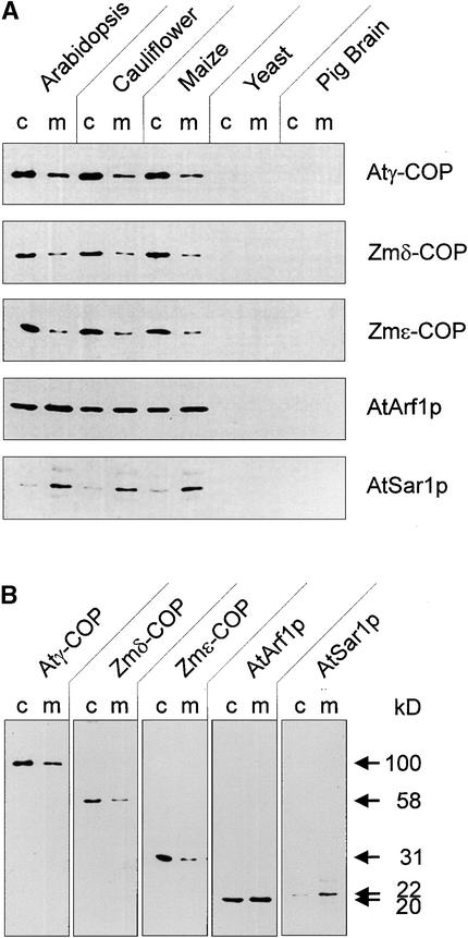
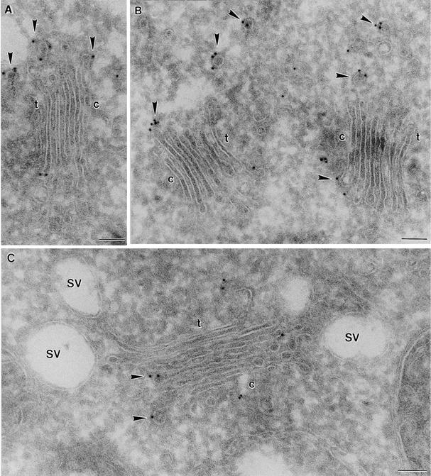
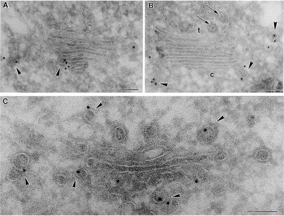
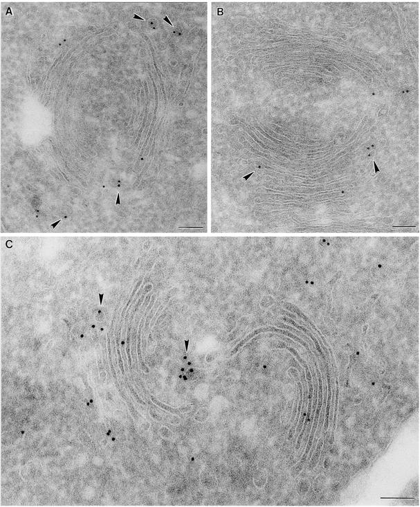
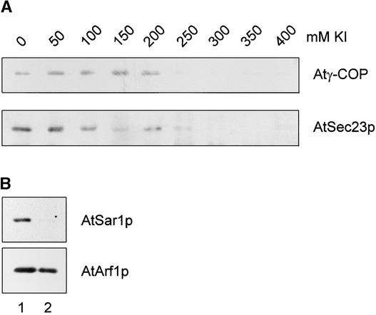
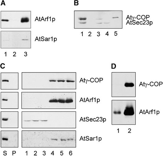
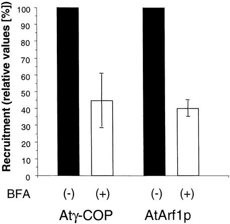
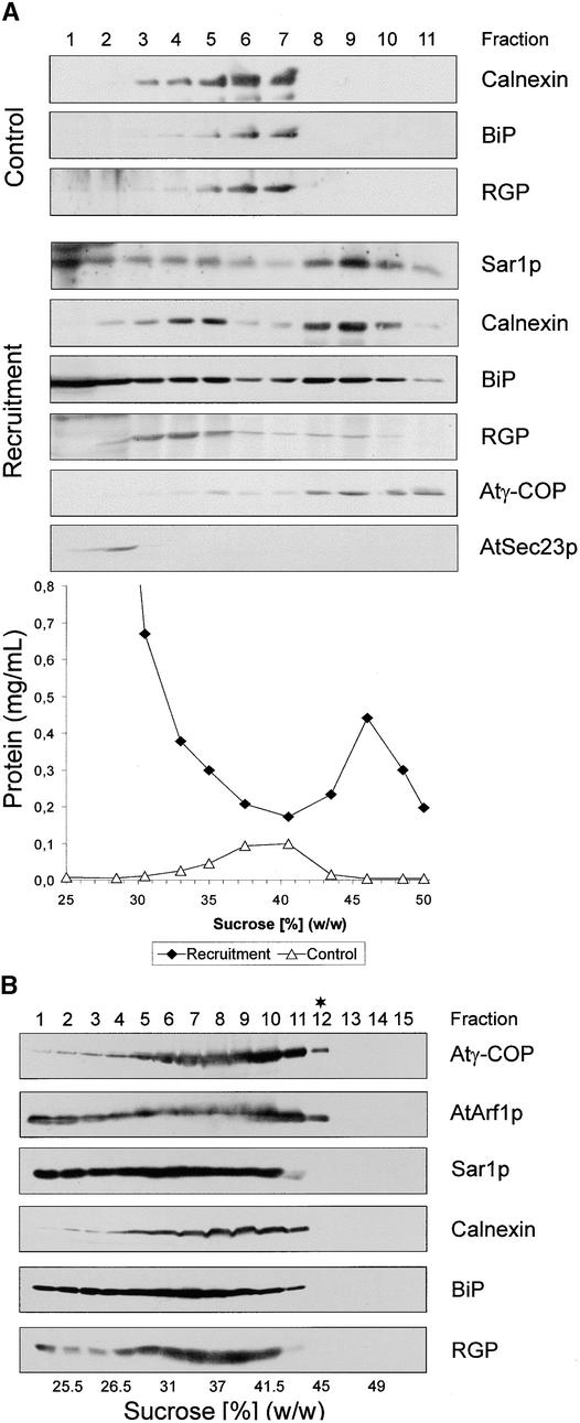
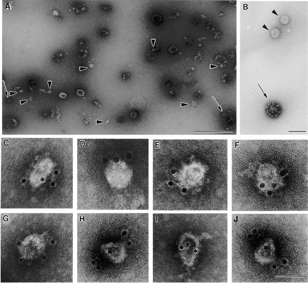
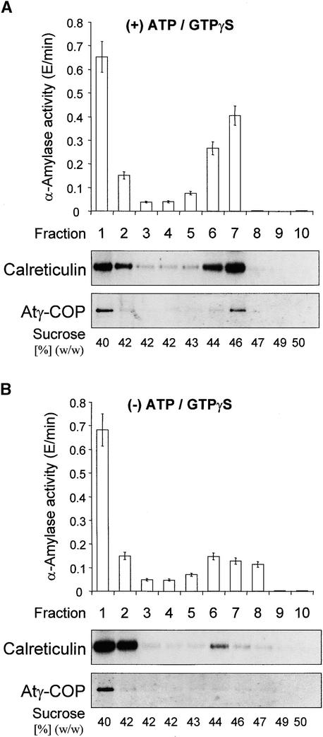
Coat protein (COP)-coated vesicles have been shown to mediate protein transport through early steps of the secretory pathway in yeast and mammalian cells. Here, we attempt to elucidate their role in vesicular trafficking of plant cells, using a combined biochemical and ultrastructural approach. Immunogold labeling of cryosections revealed that COPI proteins are localized to microvesicles surrounding or budding from the Golgi apparatus. COPI-coated buds primarily reside on the cis-face of the Golgi stack. In addition, COPI and Arf1p show predominant labeling of the cis-Golgi stack, gradually diminishing toward the trans-Golgi stack. In vitro COPI-coated vesicle induction experiments demonstrated that Arf1p as well as coatomer could be recruited from cauliflower cytosol onto mixed endoplasmic reticulum (ER)/Golgi membranes. Binding of Arf1p and coatomer is inhibited by brefeldin A, underlining the specificity of the recruitment mechanism. In vitro vesicle budding was confirmed by identification of COPI-coated vesicles through immunogold negative staining in a fraction purified from isopycnic sucrose gradient centrifugation. Similar in vitro induction experiments with tobacco ER/Golgi membranes prepared from transgenic plants overproducing barley alpha-amylase-HDEL yielded a COPI-coated vesicle fraction that contained alpha-amylase as well as calreticulin.


Figures

Figure 1.
Figure 1.
Cross-Reactivities of Antisera Raised against Recombinant Proteins Prepared from cDNA Clones for Atγ-, Zmδ-, and ɛ-COP, AtARF1p, and AtSar1p. (A) Cytosolic proteins and total membranes from the organisms indicated were prepared as described in Methods and probed with the above antisera. Equal amounts of protein were added to each lane (30 μg). c, cytosol; m, membrane. (B) Complete lanes and molecular masses of the Arabidopsis cytosol and membranes indicate specific recognition of a single antigen of the corresponding size for each of the above-listed antisera.
Figure 2.
Figure 2.
Immunogold Labeling of Cryosections with Plant COP Antibodies I. Shown is the distribution of Atγ-COP homolog in maize roots. (A) and (B) Golgi stacks from cells near the meristem. (C) Golgi stack from a root cap cell. Arrowheads point to labeled budding and released COPI-coated vesicles. c, cis-face of a Golgi stack; t, trans-face of a Golgi stack; SV, slime-containing secretory vesicle. formula image.
Figure 3.
Figure 3.
Immunogold Labeling of Cryosections with Plant COP Antibodies II. Shown is labeling of maize root Golgi stacks from cells near the meristem. (A) and (B) Labeling with Zmδ- and Zmɛ-COP antibodies, respectively. (C) Labeling with AtArf1p antibodies. Arrows point to clathrin-coated vesicles; arrowheads indicate budding COPI-coated vesicles. c, cis-face of a Golgi stack; t, trans-face of a Golgi stack. formula image.
Figure 4.
Figure 4.
Immunogold Labeling of Cryosections with Plant COP Antibodies III. Shown is labeling of Arabidopsis root Golgi stacks. (A) Labeling with Atγ-COP antibodies. (B) Labeling with Zmδ-COP antibodies. (C) Labeling with Zmɛ-COP antibodies. Arrowheads point to budding COPI-coated vesicles. formula image.
Figure 5.
Figure 5.
KI Dissociation of COPI- and COPII-Coat Proteins from Cauliflower Inflorescence Membranes. (A) A Golgi-enriched fraction was treated with increasing concentrations of KI, as described in Methods, and then centrifuged to separate the membranes from the extraction medium. The pelleted membranes were then probed for the presence of coat proteins. (B) Golgi-enriched membranes were treated as in (A), but with 300 mM KI, and then were probed with GTPase antisera. Lane 1, native membranes; lane 2, KI-stripped membranes.
Figure 6.
Figure 6.
In Vitro Recruitment of Cytosolic COPI-Vesicle Coat Proteins onto KI-Stripped Membranes. (A) Recruitment of GTPases onto cauliflower membranes. KI-stripped membranes were incubated with cytosol, ATP-regenerating system, and GTPγS for 25 min at 20°C. Membranes with bound proteins were separated from unbound proteins by centrifugation. Small GTPases in each fraction were detected in a protein gel blot with specific antibodies against AtArf1p and AtSar1p. Lane 1 contains KI-stripped membranes before incubation with cytosolic proteins; lane 2, the supernatant after recruitment; and lane 3, the pelleted membranes after recruitment. (B) Selective recruitment of coatomer onto cauliflower membranes. KI-stripped membranes were incubated as described in (A), and the sediment and supernatant proteins were screened by protein gel blotting with Atγ-COP and AtSec23p antibodies. Lane 1 contains native membranes; lane 2, KI-stripped membranes; lane 3, cytosolic proteins; lane 4, supernatant after recruitment; and lane 5, pelleted membranes after recruitment. (C) Absence of precipitable Atγ-COP, AtArf1p, AtSec23p, and AtSar1p in cytosol only (left). Nonsaturation of coat protein recruitments onto cauliflower membranes (right). The ratio of cytosolic:membrane proteins in the recruitment assay was increased by 30% (lanes 1 and 4), 100% (lanes 2 and 5), and 200% (lanes 3 and 6) over the standard values (6 mg mL−1:0.2 mg L−1). Lanes 1 to 3 represent the supernatant fractions after recruitment; lanes 4 to 6, the corresponding pelleted membrane fractions. S represents the supernatant, P the pellet after recruitment in the absence of membranes. Membranes, medium, incubation, and other conditions were as described in (A). Antibody screening was as described in (A) and (B). (D) Heterologous recruitment of COPI-vesicle coat proteins from cauliflower cytosol onto Golgi/ER membranes from tobacco mesophyll. Tobacco membranes were isolated as described in Methods, incubated with cauliflower cytosol, and subsequently screened with antibodies under the conditions used in (A). Lane 1 contains KI-stripped membranes; lane 2, pelleted membranes after recruitment.
Figure 7.
Figure 7.
BFA Inhibits Recruitment of Coatomer and AtArf1p in Cauliflower Recruitment Assay. Bound coatomer/Arf1p in control and BFA-treated samples was quantified by densitometry of protein gel blots of samples of equal volume. Shown are the results of four separate experiments whose average is given as columns; the error bars represent the extent of the variation between experiments. (+) denotes the presence of 300 μM BFA and (−) the absence of BFA in the incubation media.
Figure 8.
Figure 8.
Detection of in Vitro–Formed COPI-Coated Vesicles. (A) Recruitment of coatomer onto KI-stripped membranes leads to an increase in their equilibrium density. Stripped membranes from cauliflower inflorescence were incubated in the absence or presence of cauliflower cytosolic proteins, as described for Figure 6A. The incubation mixtures were then layered onto sucrose density gradients and centrifuged to equilibrium, as described in Methods. Fractions were screened with antibodies against ER (calnexin, BiP), Golgi (RGP), and COPI/COPII coat proteins (Atγ-COP, AtSec23p). As depicted, stripped membranes banded at densities ∼10% (w/w) less than those at which the recruited membranes did. (B) Isolation of COPI-coated vesicles. KI-stripped cauliflower membranes were incubated with cauliflower cytosol under standard conditions, extracted with high-salt reagent (250 mM KCl) for 30 min at 4°C, and centrifuged at 30,000g; the membranes in the supernatant were subjected to isopcynic sucrose density gradient centrifugation as in (A). Equal-volume fractions were then monitored in protein gel blots with the antisera described above. The asterisk denotes the putative COPI-coated vesicle–containing fraction used for Figure 9.
Figure 9.
Figure 9.
Immunogold Negative Staining of the Contents of Fraction 12 from the Gradient Shown in Figure 8B. (A) Overview of normal negative staining. Putative COPI-coated vesicles are indicated with arrowheads, clathrin-coated vesicles with arrows. (B) Comparison of clathrin- and COP-coated vesicles. Arrows and arrowheads are as in (A). (C) to (J) Gallery of negatively stained putative COPI-coated vesicles decorated with gold-coupled Zmɛ-COP antibodies. formula image; formula image; formula image.
Figure 10.
Figure 10.
Heterologous in Vitro Induction of COPI-Coated Vesicles from Cauliflower Cytosol and Tobacco Membranes from Plants Expressing α-Amylase–HDEL. (A) Incubation performed in the presence (+) of ATP/GTPγS. The histogram represents the profile of α-amylase activity. The protein gel blots of the corresponding fractions document the distributions of calreticulin and Atγ-COP in relation to α-amylase activity. (B) Incubation performed in the absence (−) of ATP/GTPγS. All incubations and membrane separations were as given in Figure 8B. Activity measurements were performed in triplicate; error bars represent the extent of variation.

Comment in

References

    1. Andreeva, A.V., Kutuzov, M.A., Evans, D.E., and Hawes, C.R. (1998). The structure and function of the Golgi apparatus: A hundred years of questions. J. Exp. Bot. 49, 1281–1291.
    1. Aridor, M., Bannykh, S.I., Rowe, T., and Balch, W.E. (1995). Sequential coupling between COPII and COPI vesicle coats in endoplasmic reticulum to Golgi transport. J. Cell Biol. 131, 875–893. - PMC - PubMed
    1. Aridor, M., Weissman, J., Bannykh, S.I., Nuoffer, C., and Balch, W.E. (1998). Cargo selection by the COPII budding machinery during export from the endoplasmic reticulum. J. Cell Biol. 141, 61–70. - PMC - PubMed
    1. Bannykh, S.I., and Balch, W.E. (1998). Selective transport of cargo between the endoplasmic reticulum and Golgi compartments. Histochem. Cell Biol. 109, 463–475. - PubMed
    1. Barlowe, C., and Schekman, R. (1993). SEC12 encodes a guanine nucleotide exchange factor essential for transport vesicle formation from the ER. Nature 365, 347–349. - PubMed

Publication types

LinkOut - more resources