Surgical correction for lower lid epiblepharon in Asians
- PMID: 11090483
- PMCID: PMC1723336
- DOI: 10.1136/bjo.84.12.1407
Surgical correction for lower lid epiblepharon in Asians
Abstract
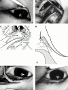
Background/aims: Epiblepharon is a congenital lid anomaly in which a fold of skin and underlying orbicularis muscle push the lashes against the eyeball. It is important to get a good lash eversion effect without forming a prominent lid crease in Asian patients. The surgical effect of this rotating suture technique was evaluated.
Methods: Surgical correction for epiblepharon was performed on 197 patients and the results analysed in 169 patients who had been followed for 1 month or more. After subciliary incision, several buried 8-0 nylon sutures were placed to allow adhesion between the tarsal plate and the subcutaneous tissue of the upper skin flap with minimal resection of pretarsal orbicularis and redundant skin.
Results: 156 patients (92.3%) showed satisfactory results during 7.1 months of average follow up. Reoperation was performed only on two patients out of 13 because of mildness of symptoms and signs. Complications were minimal including suture abscesses in four patients and wound dehiscence in one.
Conclusion: The rotating suture technique was very effective in repairing epiblepharon without forming a prominent lower eyelid crease.
Figures


References
MeSH terms
LinkOut - more resources
Full Text Sources
Medical
