. 2000 Nov;122(2):150-6.
doi: 10.1046/j.1365-2249.2000.01333.x.

The inhibition of cutaneous T cell apoptosis may prevent resolution of inflammation in atopic eczema

Affiliations

The inhibition of cutaneous T cell apoptosis may prevent resolution of inflammation in atopic eczema

C H Orteu et al. Clin Exp Immunol. 2000 Nov.

Abstract

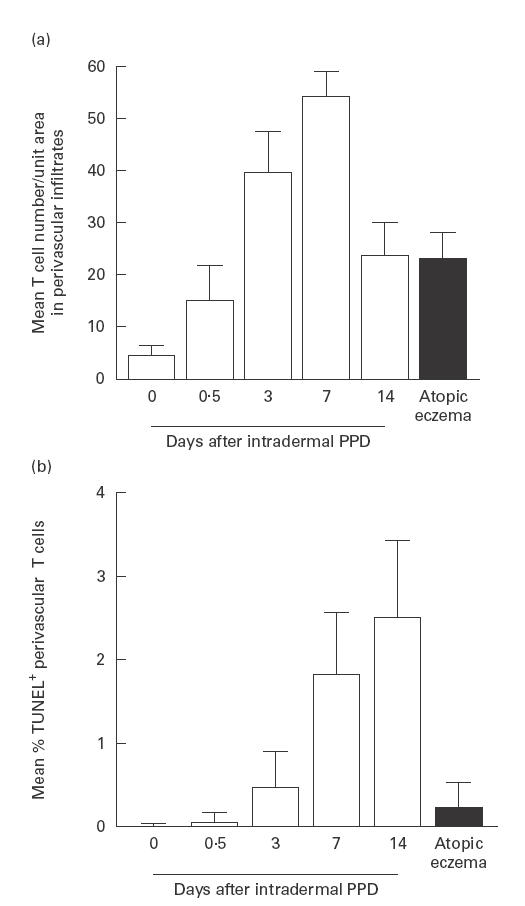
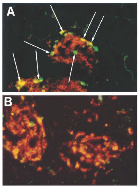
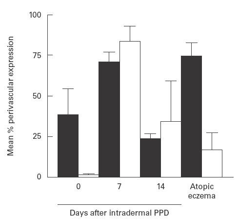
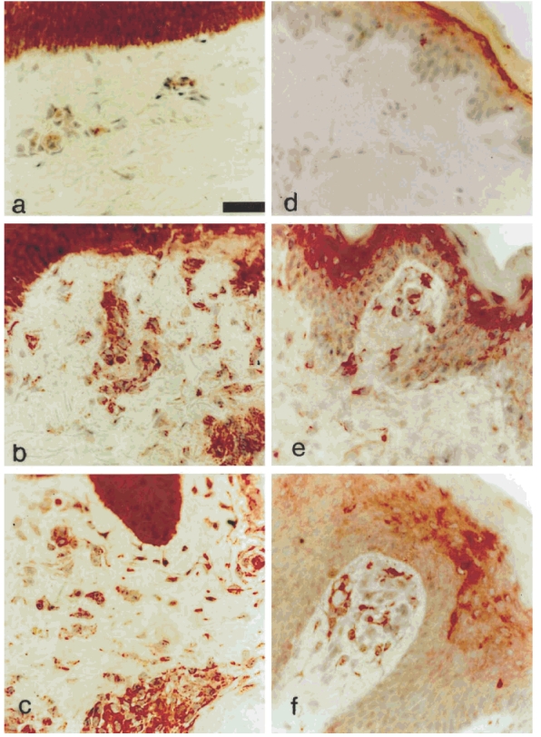
Atopic eczema (AE) is characterized by the persistence of infiltrating T lymphocytes in the dermis. To test the hypothesis that dysregulation of normal T cell apoptosis may contribute to the pathogenesis and chronicity of AE we compared patients with a normal resolving immune response (Mantoux reaction (MR)) induced in healthy volunteers by cutaneous PPD injection. Significantly less T cell apoptosis was observed in lesional skin of AE patients compared with either the peak or the resolution phase of the MR (P < 0.0001). The low incidence of T cell apoptosis in AE was associated with significantly increased levels of Bcl-2 relative to Bax (P < 0.0001) and significantly decreased CD95-L expression (P < 0.002) compared with the resolving MR. The cytokines IL-15 and interferon-beta (IFN-beta), which prevent activated T cell apoptosis, were expressed maximally on day 7 and day 14 of the MR, respectively. In contrast, AE patients expressed high levels of both IL-15 and IFN-beta in cutaneous lesions at the same time. This suggests that the co-expression of two anti-apoptotic cytokines, which are not found together during resolving cutaneous responses, may contribute to excessive T cell survival which leads to the persistence of inflammation in patients with AE.


Figures

Fig. 1
Fig. 1
Perivascular T cell numbers in normal skin, Mantoux reactions (MR) and chronic atopic eczema (AE). (a) T cells were stained by an indirect immunoperoxidase method and quantified in each section using an image analysis system as described in PATIENTS and METHODS (normal skin n = 5; MR, days 0·5, 3 and 14, n = 5, day 7 n = 4; AE, n = 9). Error bars indicate s.d. (b) Apoptosis was determined by terminal deoxynucleotidyl transferase-mediated dUTP-biotin nick end labelling (TUNEL) staining in normal skin, MR and chronic AE. Dual immunofluorescence (IMF) was used to count proportions of perivascular T cells expressing TUNEL reactivity (normal skin, days 7 and 14 MR, n = 4, day 0·5 and 3 MR, n = 5; AE n = 9). Cells were quantified using a Zeiss fluorescence microscope as described in PATIENTS and METHODS. Error bars indicate s.d.
Fig. 2
Fig. 2
The incidence of T cell apoptosis within perivascular infiltrates during the resolution phase (day 14) of the Mantoux reaction (MR) ((A) see arrows) and in a patient with atopic eczema (AE) (B) was determined by two-colour immunofluorescence staining (see legend to Fig. 1). T cells (orange) and terminal deoxynucleotidyl transferase-mediated dUTP-biotin nick end labelling (TUNEL) (green) staining were quantified using a Zeiss fluorescence microscope as described in Patients and Methods.
Fig. 3
Fig. 3
T cell Bcl-2 (▪) and CD95 ligand expression (□) in perivascular infiltrates in normal skin, Mantoux reactions (MR) and chronic atopic eczema (AE). Dual immunofluorescence studies were performed and the proportion of T cells which also expressed Bcl-2 was quantified using a Zeiss fluorescence microscope as described in PATIENTS and METHODS. A biotin/streptavidin method and an image analysis system were used to determine the proportion of perivascular cells expressing CD95 ligand. Cells with cytoplasmic or membrane positivity were counted (normal skin and day 7 MR, n = 4; day 14 MR, n = 5; AE, n = 8). Error bars indicate s.d.
Fig. 4
Fig. 4
Photomicrographs stained by means of biotin/streptavidin technique with MoAb in skin sections. Staining with anti-IL-15 MoAb (a–c) shows strongly positive keratinocytes, but little dermal staining in normal skin (a), strong positivity in keratinocytes, perivascular areas and interstitial cells on day 14 of the Mantoux reaction (MR) (b). Similar strong positivity is observed in keratinocytes, interstitial (predominantly dendritic) cells and perivascular areas in atopic eczema (AE) (c). Staining with anti-IFN-β antibody (d–f) shows weak suprabasal keratinocyte positivity in normal skin (d), and increased positivity in keratinocytes and positive dendritic cells in the papillary dermis on day 3 of the MR (e). In AE weak–moderate keratinocyte positivity, and numerous strongly positive dendritic and perivascular cells in the papillary dermis are present. (Original mag. × 400, calibration bar 24 μ m.)

References

    1. Krammer PH, Behrmann I, Daniel P, Dhein J, Debatin KM. Regulation of apoptosis in the immune system. Curr Opin Immunol. 1994;6:279–89. - PubMed
    1. Akbar AN, Salmon M. Cellular environments and apoptosis: tissue microenvironments control activated T cell death. Immunol Today. 1997;18:72–76. - PubMed
    1. Salmon M, Scheel-Toellner D, Huissoon AP, et al. Inhibition of T cell apoptosis in the rheumatoid synovium. J Clin Invest. 1997;99:439–46. - PMC - PubMed
    1. Pilling D, Akbar AN, Girdlestone J, et al. Interferon-beta mediates stromal cell rescue of T cells from apoptosis. Eur J Immunol. 1999;29:1041–50. - PubMed
    1. Salmon M, Pilling D, Borthwick NJ, Akbar AN. Inhibition of T cell apoptosis: a mechanism for the persistence of chronic inflammation. Immunologist. 1997;5:87–92.

Publication types