

Th1 cytokine pattern in sarcoidosis is expressed by bronchoalveolar CD4+ and CD8+ T cells
- PMID: 11091281
- PMCID: PMC1905777
- DOI: 10.1046/j.1365-2249.2000.01365.x
Th1 cytokine pattern in sarcoidosis is expressed by bronchoalveolar CD4+ and CD8+ T cells
Abstract
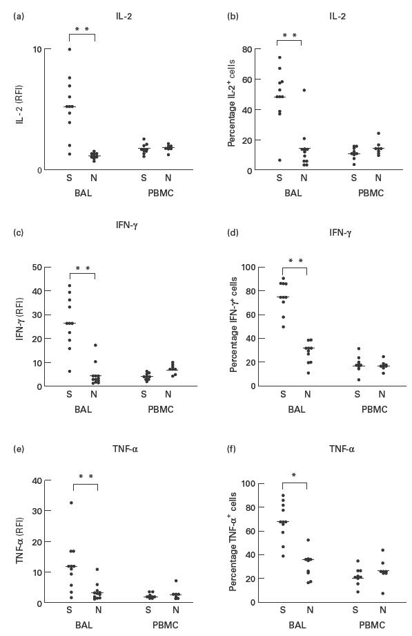
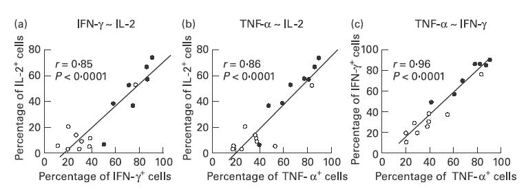
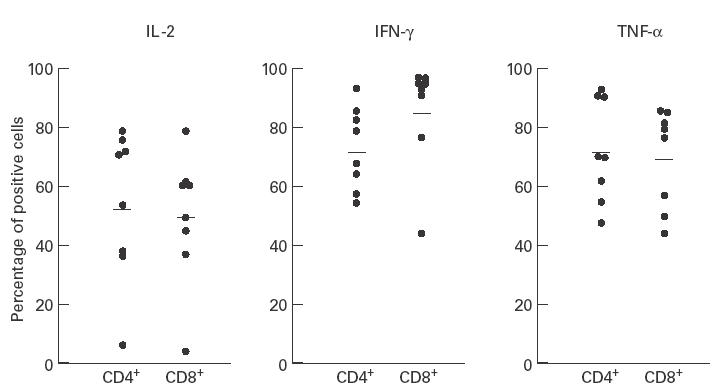
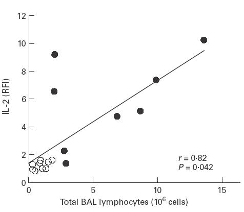
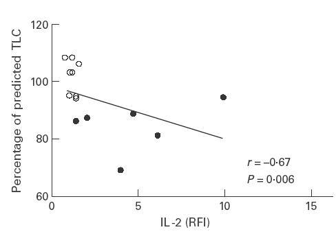
The pathogenesis of pulmonary sarcoidosis has been related to an increased production of Th1-like cytokines. However, cytokine expression in sarcoidosis has not been systematically studied at a single-cell level. We therefore investigated the expression of IL-2, IL-4, IL-13, tumour necrosis factor-alpha (TNF-alpha) and interferon-gamma (IFN-gamma) intracellularly in bronchoalveolar lavage (BAL) and peripheral blood CD3+ T lymphocytes from patients with pulmonary sarcoidosis (radiologic stage II-III, n = 8) and normal controls (n = 9) by flow cytometry. In contrast to IL-4 and IL-13, the percentage of T lymphocytes expressing intracellular IL-2 (49.3 +/- 21.3% versus 14.5 +/- 15.6%), IFN-gamma (75.5 +/- 14.9% versus 32.6 +/- 18.7%) and TNF-alpha (68.3 +/- 18.7% versus 36.8 +/- 20.8%) was significantly higher in patients with sarcoidosis than in normal controls (each P < 0.005). In contrast to BAL lymphocytes, expression of these cytokines in peripheral blood lymphocytes did not differ between patients with sarcoidosis and normal controls. Close correlations were observed between the percentages of BAL lymphocytes expressing intracellular IL-2, IFN-gamma and TNF-alpha, but not for IL-4 or IL-13. Analysis of the expression of these cytokines in T lymphocyte subsets revealed IL-2, IFN-gamma, and TNF-alpha in CD4+ as well as CD8+ T lymphocytes, suggesting a contribution of TC1 cells to the production of proinflammatory cytokines in sarcoidosis. We conclude that a Th1-like cytokine pattern can be observed in CD4+ as well as in CD8+ BAL T lymphocytes in patients with pulmonary sarcoidosis.
Figures
References
-
- Hunninghake GW, Crystal RG. Pulmonary sarcoidosis: a disorder mediated by excess helper T-lymphocyte activity at sites of disease activity. N Engl J Med. 1981;305:429–34. - PubMed
-
- Müller-Quernheim J, Saltini C, Sondermeyer P, Crystal RG. Compartmentalized activation of the interleukin 2 gene by lung T lymphocytes in active pulmonary sarcoidosis. J Immunol. 1986;137:3475–83. - PubMed
Publication types
MeSH terms
Substances
LinkOut - more resources
Full Text Sources
Other Literature Sources
Medical
Research Materials
