

Reduced expression of the complement receptor type 2 (CR2, CD21) by synovial fluid B and T lymphocytes
- PMID: 11091285
- PMCID: PMC1905766
- DOI: 10.1046/j.1365-2249.2000.01379.x
Reduced expression of the complement receptor type 2 (CR2, CD21) by synovial fluid B and T lymphocytes
Abstract
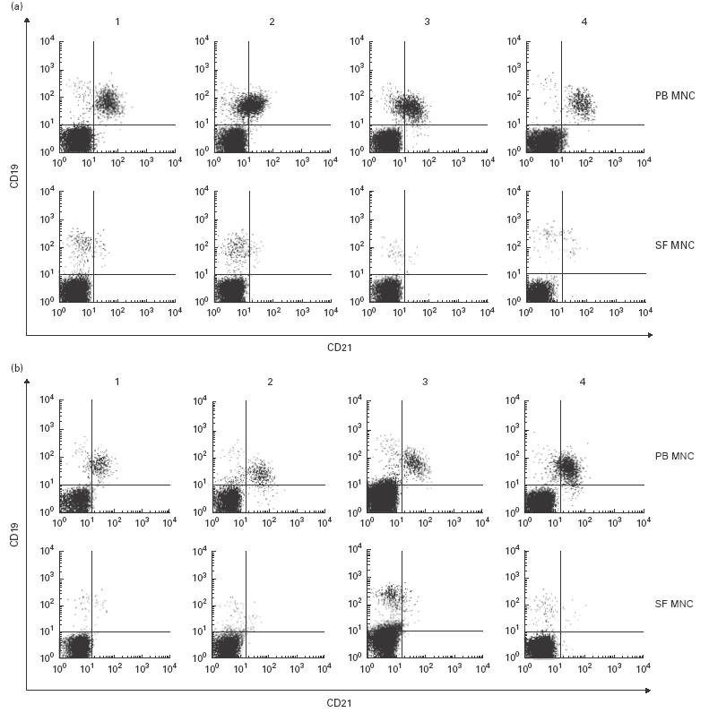
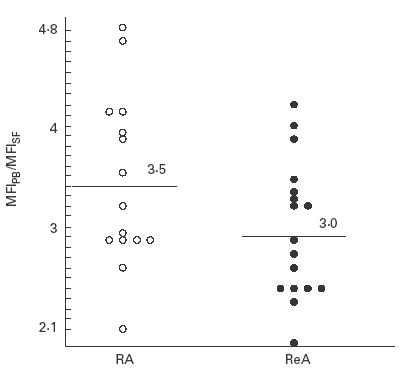
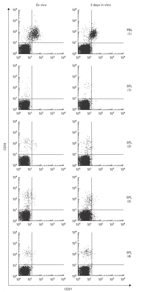
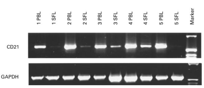
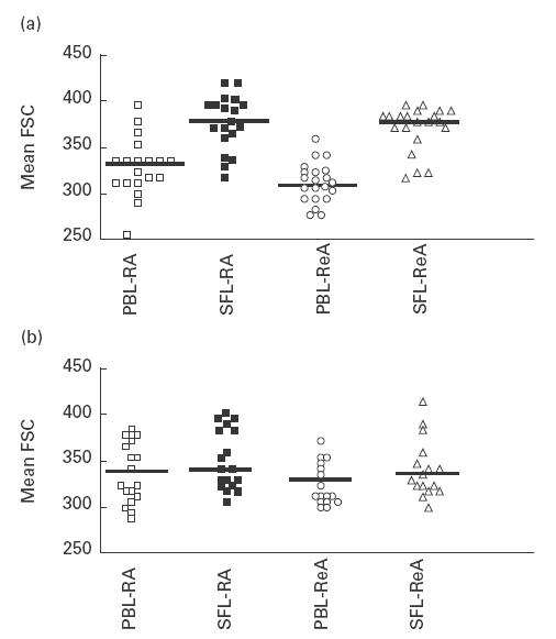
The expression of CR2 (CD21) by synovial B and T lymphocytes of patients suffering from various forms of arthritis was analysed with cytofluorometry and with reverse transcriptase-polymerase chain reaction. CR2 (CD21) cell surface protein was detected in normal quantities on peripheral B cells, but was almost absent on synovial B lymphocytes of the same patients. This reduction was most severe in patients with rheumatoid arthritis, but also observed in all other cases. CR2 (CD21) did not reappear after in vitro culture. CR2 (CD21) mRNA was also strongly reduced in synovial B and T lymphocytes. Synovial fluid B lymphocytes were larger than peripheral blood B lymphocytes, while T cells from the same patients showed no size differences. We conclude that synovial fluid B lymphocytes have undergone an irreversible step towards terminal differentiation. The presence or absence of CR2 (CD21) mRNA in peripheral versus synovial T cells indicates that CR2 (CD21) is also differentially expressed by T lymphocytes.
Figures
Similar articles
-
Silencing of CD21 expression in synovial lymphocytes is independent of methylation of the CD21 promoter CpG island.Rheumatol Int. 2001 May;20(4):133-7. doi: 10.1007/s002960000090. Rheumatol Int. 2001. PMID: 11411956
-
Human B and T lymphocytes have similar amounts of CD21 mRNA, but differ in surface expression of the CD21 glycoprotein.Int Immunol. 1998 Aug;10(8):1197-202. doi: 10.1093/intimm/10.8.1197. Int Immunol. 1998. PMID: 9723706
-
Lymphocytes in rheumatoid and nonrheumatoid synovial fluids. Nonspecificity of high T-cell and low B-cell percentages.Ann Rheum Dis. 1975 Oct;35(5):451-5. doi: 10.1136/ard.35.5.451. Ann Rheum Dis. 1975. PMID: 1086654 Free PMC article.
-
Expression and role of CR1 and CR2 on B and T lymphocytes under physiological and autoimmune conditions.Mol Immunol. 2009 Sep;46(14):2767-73. doi: 10.1016/j.molimm.2009.05.181. Epub 2009 Jun 25. Mol Immunol. 2009. PMID: 19559484 Review.
-
B lymphocyte function in patients with rheumatoid arthritis: impact of regulatory T lymphocytes and macrophages--modulation by antirheumatic drugs.Dan Med Bull. 1988 Apr;35(2):140-57. Dan Med Bull. 1988. PMID: 3282810 Review.
Cited by
-
Decreased levels of sCD21 and sCD23 in blood of patients with systemic-juvenile arthritis, polyarticular-juvenile arthritis, and pauciarticular-juvenile arthritis.Rheumatol Int. 2012 Jun;32(6):1581-7. doi: 10.1007/s00296-011-1830-1. Epub 2011 Feb 17. Rheumatol Int. 2012. PMID: 21328056
-
Expansion of Activated Peripheral Blood Memory B Cells in Rheumatoid Arthritis, Impact of B Cell Depletion Therapy, and Biomarkers of Response.PLoS One. 2015 Jun 5;10(6):e0128269. doi: 10.1371/journal.pone.0128269. eCollection 2015. PLoS One. 2015. PMID: 26047509 Free PMC article.
-
Cytometric profiling in various clinical forms of multiple sclerosis with respect to CD21+, CD32+, and CD35+ B and T cells.Transl Neurodegener. 2013 Jul 2;2(1):14. doi: 10.1186/2047-9158-2-14. Transl Neurodegener. 2013. PMID: 23819946 Free PMC article.
-
Soluble CD21 in sera and synovial fluid of arthritic patients.Rheumatol Int. 2006 Jan;26(3):240-3. doi: 10.1007/s00296-004-0541-2. Epub 2005 Mar 16. Rheumatol Int. 2006. PMID: 15770483
-
The human complement receptor type 2 (CR2)/CR1 fusion protein TT32, a novel targeted inhibitor of the classical and alternative pathway C3 convertases, prevents arthritis in active immunization and passive transfer mouse models.Mol Immunol. 2019 Jan;105:150-164. doi: 10.1016/j.molimm.2018.09.013. Epub 2018 Dec 1. Mol Immunol. 2019. PMID: 30513451 Free PMC article.
References
-
- Feldmann M, Brennan FM, Maini RN. Rheumatoid arthritis. Cell. 1996;85:307–10. - PubMed
-
- Arnett FC, Edworthy SM, Block DA, et al. The American Rheumatism Association 1987 revised criteria for the classification of rheumatoid arthritis. Arthritis Rheum. 1988;31:315–24. - PubMed
-
- Zvaifler NJ, Firestein GS. Pannus and pannocytes. Alternative models of joint destruction in rheumatoid arthritis. Arthritis Rheum. 1994;37:783–9. - PubMed
-
- Folkman J. Angiogenesis in cancer, vascular, rheumatoid and other disease. Nature Med. 1995;1:27–31. - PubMed
Publication types
MeSH terms
Substances
LinkOut - more resources
Full Text Sources
Medical
