. 2000 Dec;182(24):6927-32.
doi: 10.1128/JB.182.24.6927-6932.2000.

Pleiotropic effect of protein P6 on the viral cycle of bacteriophage phi29

Affiliations

Pleiotropic effect of protein P6 on the viral cycle of bacteriophage phi29

A Camacho et al. J Bacteriol. 2000 Dec.

Abstract

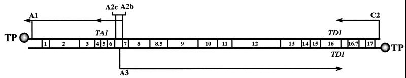
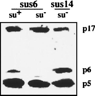
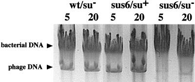
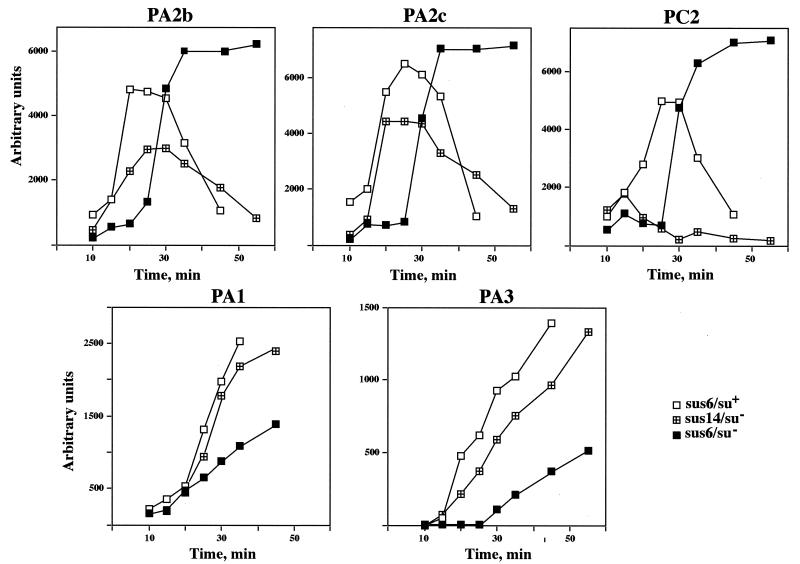
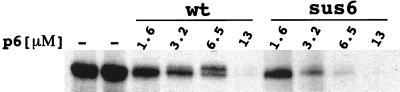
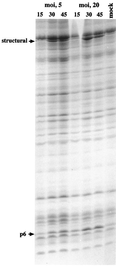
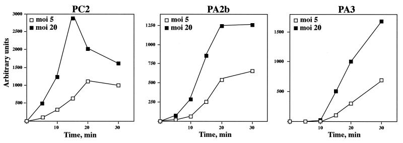
The product of bacteriophage phi29 early gene 6, protein p6, is a double-stranded-DNA binding protein and one of the more abundant proteins during viral infection. We have studied the role of protein p6 in vivo through the infection of suppressor and nonsuppressor Bacillus subtilis strains with a phage carrying a nonsense mutation in gene 6, sus6(626). In the absence of functional protein p6, the two major processes of the viral cycle, transcription and DNA replication, were affected. Viral DNA synthesis was practically abolished, and early transcription was remarkably delayed and, in addition, underregulated at late times of the infection. The amount of protein p6 synthesized after infection with mutant phage sus6(626) under suppressor conditions was sixfold lower than that produced after wild-type infection. Nonetheless, phage production was as high as that obtained after wild-type infection. These results indicate that p6 is synthesized in amounts higher than those needed for most of its functions. However, the concentration of protein p6 appeared to be important for repression of the early promoter C2.


Figures

FIG. 1
FIG. 1
Transcription map of the bacteriophage φ29 genome. Locations of promoters A1, A2c, A2b, A3, and C2 are indicated by vertical bars. Arrows indicate the direction of transcription, with the arrowheads at the termination sites (TA1 and TD1). The genetic map is depicted. The phage terminal protein (TP) is shown attached to the 5′ ends of the genome.
FIG. 2
FIG. 2
Analysis of protein p6 in B. subtilis infected with mutant sus6(626). Polypeptides present 40 min after the infection of suppressor (su+) and nonsuppressor (su) B. subtilis with φ29 mutants carrying nonsense mutations in gene 6 (sus6) and gene 14 (sus14) were separated by electrophoresis in a 10-to-20% gradient polyacrylamide gel in the presence of SDS. Proteins were transferred to a Millipore polyvinylidene difluoride membrane, and proteins p5, p6, and p17 were detected with a mixture of the corresponding monospecific antisera.
FIG. 3
FIG. 3
DNA synthesis in B. subtilis infected with mutant sus6(626). Agarose electrophoresis of the DNA present after 60 min of infection of nonsuppressor (su) and suppressor (su+) B. subtilis with φ29 mutants carrying nonsense mutations in gene 6 (sus6) or gene 14 (sus14). The MOI (phage per bacterium) is indicated on top of each lane.
FIG. 4
FIG. 4
Primer extension analysis of the transcripts produced throughout the infection cycle in B. subtilis infected with mutant sus6(626). RNA synthesized by mutant sus6(626) in nonsuppressor (su) or suppressor (su+) B. subtilis or by the control mutant sus14(1242) at an MOI of 5 was isolated at the time after the infection indicated at the bottom of each graph. Transcripts derived from each of the promoters (indicated above each graph) were analyzed by the extension of radioactively labeled specific primers and quantitated as described in Materials and Methods. The graphs show the amount of mRNA observed during an infection cycle, where each point represents the average of at least three independent experiments. Arbitrary units are comparable between graphs.
FIG. 5
FIG. 5
Effect of protein p6 on in vitro transcription of promoter C2. Runoff assays were performed in the presence of the indicated amounts of purified protein p6 from a wild-type (wt) phage infection or purified p6 from B. subtilis su+ infected with the mutant sus6(626) (sus6).
FIG. 6
FIG. 6
Analysis of the amount of protein p6 induced as a function of the MOI with mutant sus6(626). The suppressor B. subtilis strain was infected with mutant sus6(626) at an MOI of 5 or 20, and at the times indicated (in minutes), proteins were analyzed by SDS-polyacrylamide gel electrophoresis and stained with Coomassie blue.
FIG. 7
FIG. 7
Viral RNA synthesis as a function of the MOI with mutant sus6(626). RNA derived from the early promoters C2 and A2b or the late promoter A3 was analyzed by the extension of specific primers hybridized to transcripts purified from extracts of the suppressor strain infected with mutant sus6(626) at an MOI of 5 or 20 at the indicated times.

Similar articles

Cited by

References

    1. Abril A, Salas M, Andreu J M, Hermoso J M, Rivas G. Phage φ29 protein p6 is in a monomer-dimer equilibrium that shifts to higher association states at the millimolar concentration found in vivo. Biochemistry. 1997;36:11901–11908. - PubMed
    1. Barthelemy I, Mellado R P, Salas M. In vitro transcription of bacteriophage φ29 DNA: inhibition of early promoter by the viral replication protein p6. J Virol. 1989;63:460–462. - PMC - PubMed
    1. Barthelemy I, Salas M, Mellado R P. In vivo transcription of bacteriophage φ29 DNA: transcription termination. J Virol. 1987;61:1751–1755. - PMC - PubMed
    1. Bravo A, Salas M. Initiation of bacteriophage φ29 DNA replication in vivo: assembly of a membrane-associated multiprotein complex. J Mol Biol. 1997;269:102–112. - PubMed
    1. Carrascosa J L, Camacho A, Moreno F, Jiménez F, Mellado R P, Viñuela E, Salas M. Bacillus subtilis bacteriophage φ29. Characterization of gene products and functions. Eur J Biochem. 1976;66:229–241. - PubMed

Publication types

MeSH terms