. 2000 Dec;20(24):9236-46.
doi: 10.1128/MCB.20.24.9236-9246.2000.

Rat protein tyrosine phosphatase eta suppresses the neoplastic phenotype of retrovirally transformed thyroid cells through the stabilization of p27(Kip1)

Affiliations

Rat protein tyrosine phosphatase eta suppresses the neoplastic phenotype of retrovirally transformed thyroid cells through the stabilization of p27(Kip1)

F Trapasso et al. Mol Cell Biol. 2000 Dec.

Abstract

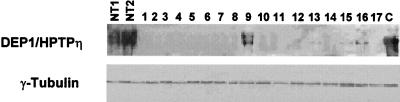
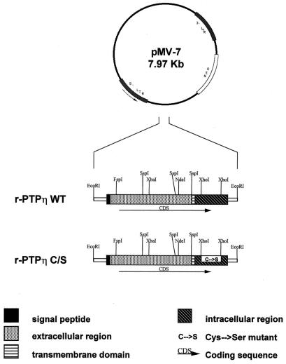
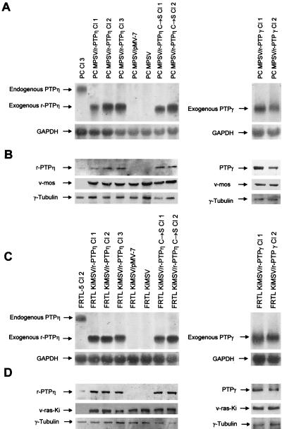
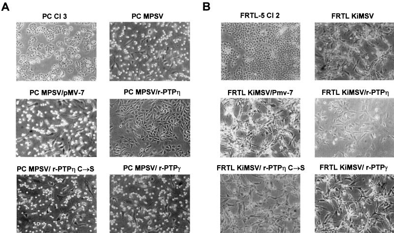
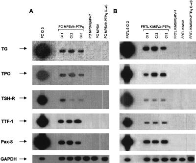
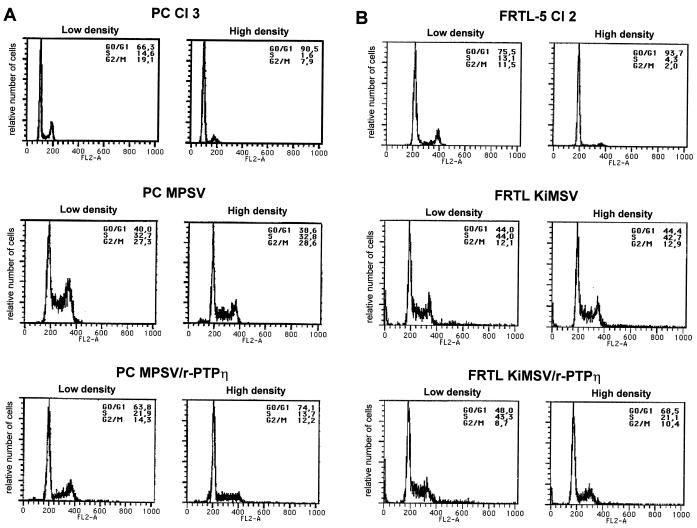
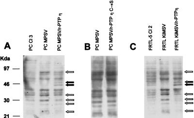
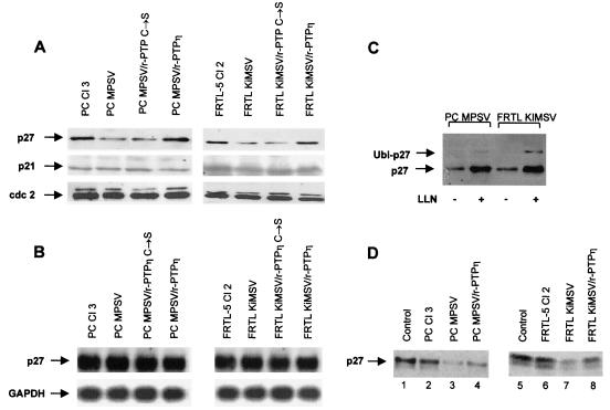
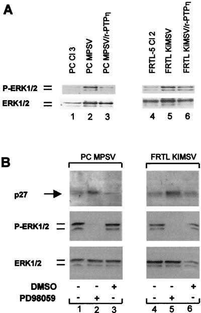
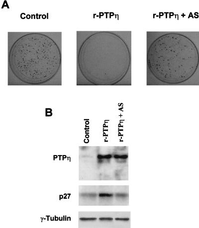
The r-PTPeta gene encodes a rat receptor-type protein tyrosine phosphatase whose expression is negatively regulated by neoplastic cell transformation. Here we first demonstrate a dramatic reduction in DEP-1/HPTPeta (the human homolog of r-PTPeta) expression in a panel of human thyroid carcinomas. Subsequently, we show that the reexpression of the r-PTPeta gene in highly malignant rat thyroid cells transformed by retroviruses carrying the v-mos and v-ras-Ki oncogenes suppresses their malignant phenotype. Cell cycle analysis demonstrated that r-PTPeta caused G(1) growth arrest and increased the cyclin-dependent kinase inhibitor p27(Kip1) protein level by reducing the proteasome-dependent degradation rate. We propose that the r-PTPeta tumor suppressor activity is mediated by p27(Kip1) protein stabilization, because suppression of p27(Kip1) protein synthesis using p27-specific antisense oligonucleotides blocked the growth-inhibitory effect induced by r-PTPeta. Furthermore, we provide evidence that in v-mos- or v-ras-Ki-transformed thyroid cells, the p27(Kip1) protein level was regulated by the mitogen-activated protein (MAP) kinase pathway and that r-PTPeta regulated p27(Kip1) stability by preventing v-mos- or v-ras-Ki-induced MAP kinase activation.


Figures

FIG. 1
FIG. 1
Expression of the DEP-1/HPTPη protein in normal and neoplastic thyroid tissues. Western blot analysis of DEP-1/HPTPη protein expression in thyroid tumor tissue. Total proteins (50 μg) were resolved by SDS–7% PAGE, transferred to nitrocellulose filters and probed with anti-DEP-1 antibodies. NT1 and NT2, two different normal thyroid tissues. Lanes 1 to 4, anaplastic carcinomas; lanes 5 to 13, papillary carcinomas; lanes 14 to 17, follicular carcinomas. C, control PC MPSV cells transfected with the r-PTPη gene. Anti-γ-tubulin antibodies were used to ensure uniform gel loading.
FIG. 2
FIG. 2
Schematic representation of the plasmids used in this study. The wild-type (WT) and mutated r-PTPη C/S cDNAs were inserted into the pMV-7 vector, which carries the gene for resistance to G418 as a selectable marker. The r-PTPη C/S construct was derived from the wild-type r-PTPη gene by coding sequence change resulting in a Cys-to-Ser mutation at position 1118 of the associated protein. This mutation abolished enzymatic activity. The domains of the r-PTPη protein are indicated. LTR, long terminal repeat; NEO, neomycin resistance gene.
FIG. 3
FIG. 3
Northern blot and Western blot analysis of the expression of the endogenous and exogenous r-PTPη gene in normal, transformed, and r-PTPη-transfected thyroid cells. (A) Northern blots. Total RNA (20 μg/lane) was extracted from PC Cl 3, PC MPSV/r-PTPη clones 1, 2, and 3, PC MPSV/pMV-7, PC MPSV, and PC MPSV/r-PTPη C/S clones 1 and 2 (left) and PC MPSV/r-PTPγ clones 1 and 2 (right) and hybridized to radiolabeled r-PTPη or GAPDH cDNAs, as indicated. (B) Western blots. Total proteins (20 μg/lane) were extracted from the cells used for panel A and hybridized with anti-r-PTPη protein, anti-v-mos, or anti-γ-tubulin antibodies. (C) Northern blots. Total RNA (20 μg/lane) was extracted from FRTL-5, FRTL-5 KiMSV/r-PTPη clones 1, 2, and 3, FRTL KIMSV, FRTL KIMSV/pMV-7, and FRTL KiMSV/r-PTPη C/S clones 1 and 2 (left) and FRTL KIMSV r-PTPη clones 1 and 2 (right) and hybridized to radiolabeled r-PTPη or GAPDH cDNAs as indicated. (D) Western blots. Total proteins (20 μg/lane) were extracted from the cells used for panel C and hybridized with anti-r-PTPη protein, anti-Ki-ras, or anti-γ-tubulin antibodies, respectively.
FIG. 4
FIG. 4
Morphology of normal, transformed, and r-PTPη-transfected thyroid cells. (A) Phase-contrast photomicrographs of normal thyroid cells (PC Cl 3), transformed thyroid cells (PC MPSV), transformed thyroid cells transfected with the wild-type r-PTPη gene (PC MPSV/r-PTPη), and transformed thyroid cells transfected with r-PTPη C/S or the r-PTPγ gene (PC MPSV/r-PTPη C/S and PC MPSV/r-PTPγ, respectively). (B) Phase-contrast photomicrographs of normal thyroid cells (FRTL-5 Cl 2), transformed thyroid cells (FRTL KiMSV), transformed thyroid cells transfected with the wild-type r-PTPη gene (FRTL KiMSV/r-PTPη), and transformed thyroid cells transfected with the mutant r-PTPη C/S or r-PTPγ gene (FRTL KiMSV/r-PTPη C/S and FRTL KiMSV/r-PTPγ, respectively). PC MPSV/pMV-7 and FRTL KiMSV/pMV-7 cell lines were obtained after transfection of transformed thyroid cells with backbone vector pMV-7. Magnification, ×150.
FIG. 5
FIG. 5
Analysis of thyroid-specific gene expression by RT-PCR in PC MPSV/r-PTPη and FRTL KiMSV/r-PTPη cells. The levels of TG, TPO, TSH-R, TTF-1, and PAX-8 mRNA were determined by RT-PCR (for details see Materials and Methods). The cDNAs were coamplified with GAPDH, as an internal control. Bands of comparable intensities, obtained by GAPDH coding sequence-specific primers, indicate comparable amplification of all samples. No bands were seen in non-reverse-transcribed RNAs, thus excluding DNA contamination (data not shown). The sources of the RNAs are indicated.
FIG. 6
FIG. 6
Flow-cytometric analysis of normal, transformed, and r-PTPη-transfected rat thyroid cells. The DNA contents of transformed and r-PTPη-transfected cells were analyzed by flow cytometry after propidium iodine staining. (A) PC Cl 3, PC MPSV, and PC MPSV/r-PTPη cells. (B) FRTL-5, FRTL KiMSV, and FRTL KiMSV/r-PTPη cells.
FIG. 7
FIG. 7
Western blot analyses of tyrosine phosphorylation patterns in normal, transformed, and r-PTPη-transfected thyroid cells. (A) Western blot analysis of PC Cl 3, PC MPSV, and PC MPSV/r-PTPη C/S cells using an antiphosphotyrosine (anti-P-Tyr)-specific antibody. (B) Western blot analysis of PC MPSV and PC MPSV/r-PTPη C/S cells with an anti-P-Tyr-specific antibody. (C) Western blot analysis of FRTL-5, FRTL KiMSV, and FRTL KiMSV/r-PTPη cells using an anti-P-Tyr-specific antibody. Total proteins extracted from cells, as indicated, were separated (40 μg/lane) by SDS-PAGE and transferred to PVDF membranes. Western blots were incubated first with antibodies against P-Tyr and then with horseradish peroxidase-conjugated secondary antibodies; the immunocomplexes were detected by enhanced chemiluminescence. As a control for equal loading, the filters were stained with Ponceau red. Open arrows, bands with decreased tyrosine phosphorylation; solid arrows, hyperphosphorylated bands.
FIG. 8
FIG. 8
Northern and Western blot analyses of p27Kip1 gene and protein expression in normal, transformed, and r-PTPη-transfected thyroid cells. (A) Western blot analysis. Proteins extracted from normal, transformed, and r-PTPη-transfected cells were separated (20 μg/lane) by SDS-PAGE, transferred to PVDF membranes, and analyzed with the indicated antibodies by Western blotting. As a control for equal loading, the blotted proteins were stained with Ponceau red. The sources of proteins are indicated. (B) Northern blot analysis. Total RNA (20 μg/lane) extracted from normal, transformed, and r-PTPη-transfected cells was hybridized to p27Kip1 radiolabeled cDNA and then to a rat GAPDH gene probe, as a control for RNA loading. The sources of RNAs are indicated. (C) Inhibition of proteasome by LLNL (LLN) stabilizes p27Kip1 protein levels. PC MPSV and FRTL KiMSV cells were treated with 50 μM LLNL proteasome inhibitor or with solvent alone (dimethyl sulfoxide) for 12 h. Cells were then lysed, and p27Kip1 expression was analyzed by Western blotting. Arrows, p27Kip1 or a monoubiquitinated form of p27 (Ubi-p27) in LLNL-treated cells. (D) In vitro p27Kip1 degradation assay. One microgram of recombinant p27Kip1 protein was incubated with 100 μg of proteasome extracts from PC Cl 3, PC MPSV, and PC MPSV/r-PTPη cells (left) or from FRTL-5 Cl 2, FRTL KiMSV, and FRTL KiMSV/r-PTPη cells (right) for 12 h and then loaded onto a 12.5% polyacrylamide gel, transferred to nitrocellulose membranes, and revealed by anti-p27Kip1 antibodies. Lane 1, the recombinant p27Kip1 protein was incubated in the absence of cell extracts. The sources of cell extracts are indicated.
FIG. 9
FIG. 9
Reduction of MAP kinase activity in r-PTPη-transfected thyroid cells results in up-regulation of p27Kip1 expression. (A) Western blot analysis of phosphorylated and total protein level of ERK1 and -2. Proteins extracted from normal, transformed, and r-PTPη transfected cells were separated (20 μg/lane) by SDS-PAGE, transferred to PVDF membranes, and analyzed with the indicated antibodies by Western blotting. (Top) the antibody used detected phosphorylated ERK1 and -2; (bottom) the antibody used detected total ERK1 and -2. (B) Inhibition of the MAP kinase pathway up-regulates p27Kip1 expression. (Top) Western blot analysis of p27Kip1 protein in untreated PC MPSV (lane 1) and FRTL KiMSV (lane 4) cells or in cells treated for 8 h with MEK inhibitor PD93059 (lanes 2 and 5, respectively) or solvent (dimethyl sulfoxide [DMSO]) alone (lanes 3 and 6, respectively); (middle) Western blot analysis of phosphorylated ERK1 and -2; (bottom) Western blot analysis of total ERK1 and -2.
FIG. 10
FIG. 10
Colony assay of r-PTPη transfection in PC MPSV cells. PC MPSV cells were transfected with a vector expressing r-PTPη cDNA in the presence of antisense oligonucleotides corresponding to the 5′ end of the p27Kip1 gene. The cells were selected for resistance to G418, and colonies were the counted after 14 days. (A) PC MPSV cells transfected with control backbone vector (left), PC MPSV cells transfected with r-PTPη (middle), and PC MPSV cells transfected with r-PTPη in the presence of antisense p27Kip1 oligonucleotides (AS) (right). (B) Western blot analysis of p27Kip1 and r-PTPη expression in PC MPSV cells transfected with the backbone vector (left lane), r-PTPη (middle lane), or r-PTPη in the presence of antisense p27Kip1 oligonucleotides (right lane). Anti-γ-tubulin antibodies were used to assure uniform loading of lanes.

Similar articles

Cited by

References

    1. Baldassarre G, Belletti B, Spiezia S, Bruni P, Trapasso F, Pentimalli F, Barone M V, Chiappetta G, Vento M T, Boccia A, Fusco A, Viglietto G. Overexpressed cyclin D3 contributes to retaining the growth inhibitor p27 in the cytoplasm of thyroid tumor cells. J Clin Investig. 1999;104:865–874. - PMC - PubMed
    1. Berlingieri M T, Manfioletti G, Santoro M, Bandiera A, Visconti R, Giancotti V, Fusco A. Inhibition of HMGI-C protein synthesis suppresses retrovirally induced neoplastic transformation of rat thyroid cells. Mol Cell Biol. 1995;15:1545–1553. - PMC - PubMed
    1. Brady-Kalnay S M, Rimm D L, Tonks N K. Receptor protein tyrosine phosphatase PTPmu associates with cadherins and catenins in vivo. J Cell Biol. 1995;130:977–986. - PMC - PubMed
    1. Brown-Shimer S, Johnson K A, Hill D E, Bruskin A M. Effect of protein tyrosine phosphatase 1B expression on transformation by the human neu oncogene. Cancer Res. 1992;52:478–482. - PubMed
    1. Coats S, Flanagan W M, Nourse J, Roberts J M. Requirement of p27Kip1 for restriction point control of the fibroblast cell cycle. Science. 1996;272:877–880. - PubMed

Publication types

MeSH terms

Substances

LinkOut - more resources