. 2000 Dec;20(24):9281-93.
doi: 10.1128/MCB.20.24.9281-9293.2000.

The MN1-TEL fusion protein, encoded by the translocation (12;22)(p13;q11) in myeloid leukemia, is a transcription factor with transforming activity

Affiliations

The MN1-TEL fusion protein, encoded by the translocation (12;22)(p13;q11) in myeloid leukemia, is a transcription factor with transforming activity

A Buijs et al. Mol Cell Biol. 2000 Dec.

Abstract

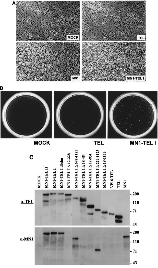
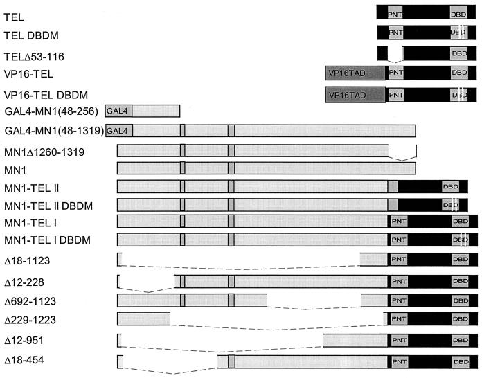
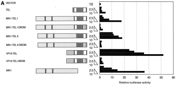
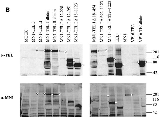
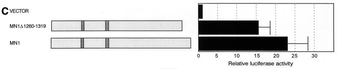
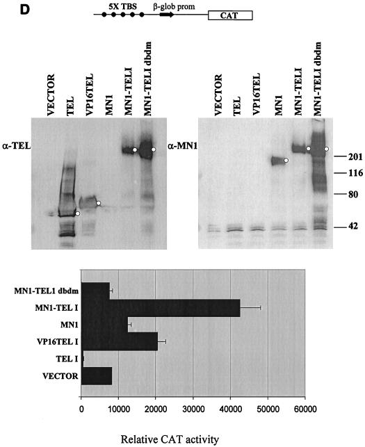
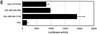
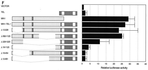
The Tel gene (or ETV6) is the target of the translocation (12;22)(p13;q11) in myeloid leukemia. TEL is a member of the ETS family of transcription factors and contains the pointed protein interaction (PNT) domain and an ETS DNA binding domain (DBD). By contrast to other chimeric proteins that contain TEL's PNT domain, such as TEL-platelet-derived growth factor beta receptor in t(5;12)(q33;p13), MN1-TEL contains the DBD of TEL. The N-terminal MN1 moiety is rich in proline residues and contains two polyglutamine stretches, suggesting that MN1-TEL may act as a deregulated transcription factor. We now show that MN1-TEL type I, unlike TEL and MN1, transforms NIH 3T3 cells. The transforming potential depends on both N-terminal MN1 sequences and a functional TEL DBD. Furthermore, we demonstrate that MN1 has transcription activity and that MN1-TEL acts as a chimeric transcription factor on the Moloney sarcoma virus long terminal repeat and a synthetic promoter containing TEL binding sites. The transactivating capacity of MN1-TEL depended on both the DBD of TEL and sequences in MN1. MN1-TEL contributes to leukemogenesis by a mechanism distinct from that of other chimeric proteins containing TEL.


Figures

FIG. 1
FIG. 1
Morphologic analysis and soft-agar assays of NIH 3T3 cells by retrovirus-transduced TEL, MN1, and MN1-TEL I. (A) Polyclonal populations of sorted CD8-positive NIH 3T3 cells mock infected or infected with TEL-, MN1-, or MN1-TEL I-expressing retroviruses were seeded on culture dishes. Only MN1-TEL I-infected NIH 3T3 cells were not contact inhibited and displayed an aberrant morphology. (B) MN1-TEL I-infected CD8-positive NIH 3T3 cells formed colonies in soft agar. Mock- or TEL-infected cells did not form colonies when plated in agar. The cells were seeded into 0.3% Noble agar at a density of 20,000 per plate and at a serum concentration of 15%. (C) Aliquots of cells transduced with retroviral vectors encoding all the different retroviral constructs (indicated above the lanes) used for soft-agar colony assays were lysed, and 50 μg of protein from each lysate was Western blotted and incubated with α-TEL antibody (top) or α-MN1 antibody (bottom).
FIG. 2
FIG. 2
Schematic representation of TEL, MN1, MN1-TEL, and VP16-TEL cDNA constructs. TAD, transactivating sequences. The white lines in DBD represent mutated codons. The gray boxes in MN1 sequences represent glutamine stretches. The dashed lines represent deleted sequences.
FIG. 3
FIG. 3
MN1 contributes transcription-activating sequences to TEL. (A) Transient-transfection experiments were performed using increasing amounts of CMV promoter-driven TEL, MN1, MN1-TEL, and VP16-TEL activator constructs (2.5 to 10 μg), as well as their respective TEL DBD mutants, with 1 μg of pMSVluc in NIH 3T3 cells. Luciferase assays were performed 24 h after removal of the calcium phosphate precipitate. The induction of luciferase (normalized to a secreted alkaline phosphatase control) is shown relative to the value of an empty vector. (B) Protein lysate (50 μg) of NIH 3T3 cells transfected in transient-transfection experiments (indicated above the lanes) was Western blotted and incubated with an α-TEL antibody (upper two blots) followed by an α-MN1 antibody (lower two blots). Bands of interest are indicated by small white circles. (C) Transient-transfection experiments using 5 μg of pCDNA3-based MN1 activator construct with 1 μg of pMSVluc in Hep3B cells. Luciferase assays were performed 40 h after removal of the calcium phosphate precipitate. The induction of luciferase is shown relative to the value of an empty vector. The mean values (+ standard deviations) of two experiments are shown. Each transfection was done in duplicate. (D) Transient-transfection experiments were performed using 1 μg of CMV promoter-driven TEL, MN1, MN1-TEL I, MN1-TEL I DBDM, and VP16-TEL activator constructs with 2 μg of 5× TEL-CAT reporter construct that contains a minimal β-globin promoter (β-glob prom) preceded by five concatemerized TEL binding sites (TBS) (CCGGAAGT) (top). In the middle is shown the relative protein expression of the different constructs used in the transient-expression assays. Each lane was loaded with 50 μg of NIH 3T3 cell lysate after transfection of each of the different effector plasmids. After Western blotting, the membrane was incubated with α-TEL antibody (left) followed by incubation with the α-MN1 antibody (right). Bands of interest are indicated by small white circles. On the bottom are shown the relative transactivations of the CAT reporter by the different constructs indicated on the left. CAT assays were performed 48 h after transfection. The values were corrected for SEAP activity derived from a cotransfected SEAP plasmid. The mean values (+ standard deviations) of four different experiments are shown. (E) Transient-transcription experiments using 0.5 μg of pCDNA3-based GAL4 DBD MN1 and VP16 fusion activator constructs with 0.5 μg of adenovirus E4 minimal-promoter-based luciferase reporter construct containing 5× GAL-responsive elements in Hep3B cells. MN1 cDNA sequences encoding amino acids 48 to 256 and 48 to 1319 or the transactivating domain of VP16 (VP16TAD) were expressed as GAL4 DBD fusion proteins. The mean values (+ standard deviations) of at least three experiments are shown. (F) Transient-transfection experiments using 3 μg of CMV promoter-driven activator constructs in NIH 3T3 cells to analyze which domains in the MN1 moiety of MN1-TEL I mediate transactivation of the MSV LTR. Normalized luciferase values relative to an empty vector are shown. The mean values (+ standard deviations) of two experiments are shown. Each transfection was performed in triplicate.
FIG. 3
FIG. 3
MN1 contributes transcription-activating sequences to TEL. (A) Transient-transfection experiments were performed using increasing amounts of CMV promoter-driven TEL, MN1, MN1-TEL, and VP16-TEL activator constructs (2.5 to 10 μg), as well as their respective TEL DBD mutants, with 1 μg of pMSVluc in NIH 3T3 cells. Luciferase assays were performed 24 h after removal of the calcium phosphate precipitate. The induction of luciferase (normalized to a secreted alkaline phosphatase control) is shown relative to the value of an empty vector. (B) Protein lysate (50 μg) of NIH 3T3 cells transfected in transient-transfection experiments (indicated above the lanes) was Western blotted and incubated with an α-TEL antibody (upper two blots) followed by an α-MN1 antibody (lower two blots). Bands of interest are indicated by small white circles. (C) Transient-transfection experiments using 5 μg of pCDNA3-based MN1 activator construct with 1 μg of pMSVluc in Hep3B cells. Luciferase assays were performed 40 h after removal of the calcium phosphate precipitate. The induction of luciferase is shown relative to the value of an empty vector. The mean values (+ standard deviations) of two experiments are shown. Each transfection was done in duplicate. (D) Transient-transfection experiments were performed using 1 μg of CMV promoter-driven TEL, MN1, MN1-TEL I, MN1-TEL I DBDM, and VP16-TEL activator constructs with 2 μg of 5× TEL-CAT reporter construct that contains a minimal β-globin promoter (β-glob prom) preceded by five concatemerized TEL binding sites (TBS) (CCGGAAGT) (top). In the middle is shown the relative protein expression of the different constructs used in the transient-expression assays. Each lane was loaded with 50 μg of NIH 3T3 cell lysate after transfection of each of the different effector plasmids. After Western blotting, the membrane was incubated with α-TEL antibody (left) followed by incubation with the α-MN1 antibody (right). Bands of interest are indicated by small white circles. On the bottom are shown the relative transactivations of the CAT reporter by the different constructs indicated on the left. CAT assays were performed 48 h after transfection. The values were corrected for SEAP activity derived from a cotransfected SEAP plasmid. The mean values (+ standard deviations) of four different experiments are shown. (E) Transient-transcription experiments using 0.5 μg of pCDNA3-based GAL4 DBD MN1 and VP16 fusion activator constructs with 0.5 μg of adenovirus E4 minimal-promoter-based luciferase reporter construct containing 5× GAL-responsive elements in Hep3B cells. MN1 cDNA sequences encoding amino acids 48 to 256 and 48 to 1319 or the transactivating domain of VP16 (VP16TAD) were expressed as GAL4 DBD fusion proteins. The mean values (+ standard deviations) of at least three experiments are shown. (F) Transient-transfection experiments using 3 μg of CMV promoter-driven activator constructs in NIH 3T3 cells to analyze which domains in the MN1 moiety of MN1-TEL I mediate transactivation of the MSV LTR. Normalized luciferase values relative to an empty vector are shown. The mean values (+ standard deviations) of two experiments are shown. Each transfection was performed in triplicate.
FIG. 4
FIG. 4
Subcellular distribution of endogenous TEL and virus-transduced TEL, MN1, MN1-TEL, and VP16-TEL proteins. (A to C) Indirect immunofluorescence analysis of endogenous TEL using immunopurified α-TEL antibodies in NIH 3T3 cells (A) and HeLa cells (B and C) and analysis of competed α-TEL antibodies using bacterially expressed glutathione S-transferase-TEL fusion protein on HeLa cells (C). (D and H) Endogenous and exogenous expression of MN1 in NIH 3T3 cells. The distributions of virus-transduced TEL, TEL DBDM, and TELΔ53-116 (E to G), MN1-TEL I and MN1-TEL I DBDM (I and J), MN1-TEL II and MN1-TEL II DBDM (K and L), deletion mutants MN1-TEL IΔ18-1123, -Δ12-228, -Δ692-1123, -Δ229-1223, -Δ18-454, and -Δ12-951 (M to R), and VP16-TEL and VP16-TEL DBDM (S and T) in NIH 3T3 cells were analyzed using α-TEL (E to G, I, J, L, and M to T) or α-MN1(D, H, and K) antibodies. Proteins were visualized with fluorescein isothiocyanate-conjugated secondary antibody. The images were obtained by using confocal microscopy. The signals of panels A to D have been electronically amplified.
FIG. 5
FIG. 5
TEL's PNT oligomerization domain is nonfunctional in MN1-TEL. (A) HeLa cells were transiently transfected with expression plasmids encoding HA1TELΔ53-116, HA1TEL, and TEL as indicated above the lanes. Proteins were immunoprecipitated with α-HA1. Complexes were separated on an SDS–10% polyacrylamide gel and electroblotted. The proteins were visualized by Western blot analysis with α-TEL antiserum. HA1TELΔ53-116, HA1TEL, and coprecipitating proteins are indicated by arrows on the right. A molecular mass standard is on the left. (B) COS-1 cells were (co)transfected with expression plasmids encoding HA1TEL or HA1TELΔ53-116 and MN1-TEL I, MN1-TEL II, and MN1-TEL IΔ229-1223 as indicated above the lanes. Following metabolic labeling with [3H]leucine, the proteins were immunoprecipitated with α-HA1 followed by immunoprecipitation with α-MN1 and analyzed on an SDS–polyacrylamide gel. The immunoprecipitates were visualized by autoradiography. HA1TEL, HA1TELΔ53-116, and coimmunoprecipitating proteins are indicated by arrows on the right. A molecular mass standard is on the left. +, present; −, absent.

Similar articles

Cited by

References

    1. Bailly R, Bosselut R, Zucman J, Cormier F, Delattre O, Roussel M, Thomas G, Ghysdael J. DNA-binding and transcriptional activation properties of the EWS-FLI-1 fusion protein resulting from the t(11;22) translocation in Ewing sarcoma. Mol Cell Biol. 1994;14:3230–3241. - PMC - PubMed
    1. Bennicelli J L, Edwards R H, Barr F G. Mechanism for transcriptional gain of function resulting from chromosomal translocation in alveolar rhabdomyosarcoma. Proc Natl Acad Sci USA. 1996;93:5455–5459. - PMC - PubMed
    1. Boskulos K E, Pognonec P, Rabault B, Begue A, Ghysdael J. Definition of an Ets1 protein domain required for nuclear localization in cells and DNA-binding activity in vitro. Mol Cell Biol. 1989;9:5718–5721. - PMC - PubMed
    1. Buijs A, Sherr S, van Baal S, van Bezouw S, van der Plas D, Geurts van Kessel A, Riegman P, Lekanne Deprez R, Zwarthoff E, Hagemeijer A, Grosveld G. Translocation (12;22)(p13;q11) in myeloproliferative disorders results in fusion of the ETS-like TEL gene on 12p13 to the MN1 gene on 22q11. Oncogene. 1995;10:1511–1519. - PubMed
    1. Carroll M, Tomasson M H, Barker G F, Golub T R, Gilliland D G. The TEL/platelet-derived growth factor β receptor (PDGFβR) fusion in chronic myelomonocytic leukemia is a transforming protein that self-associates and activates PDGFβR kinase-dependent signaling pathways. Proc Natl Acad Sci USA. 1996;93:14845–14850. - PMC - PubMed

MeSH terms

LinkOut - more resources