. 2000 Dec;20(24):9317-30.
doi: 10.1128/MCB.20.24.9317-9330.2000.

Functional interaction between the coactivator Drosophila CREB-binding protein and ASH1, a member of the trithorax group of chromatin modifiers

Affiliations

Functional interaction between the coactivator Drosophila CREB-binding protein and ASH1, a member of the trithorax group of chromatin modifiers

F Bantignies et al. Mol Cell Biol. 2000 Dec.

Abstract

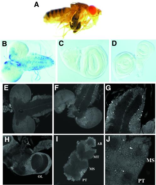
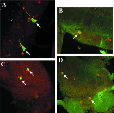
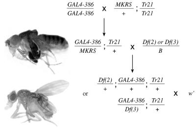
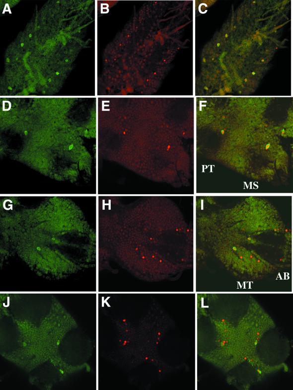
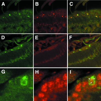
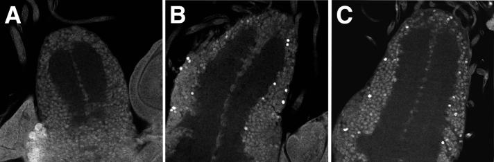
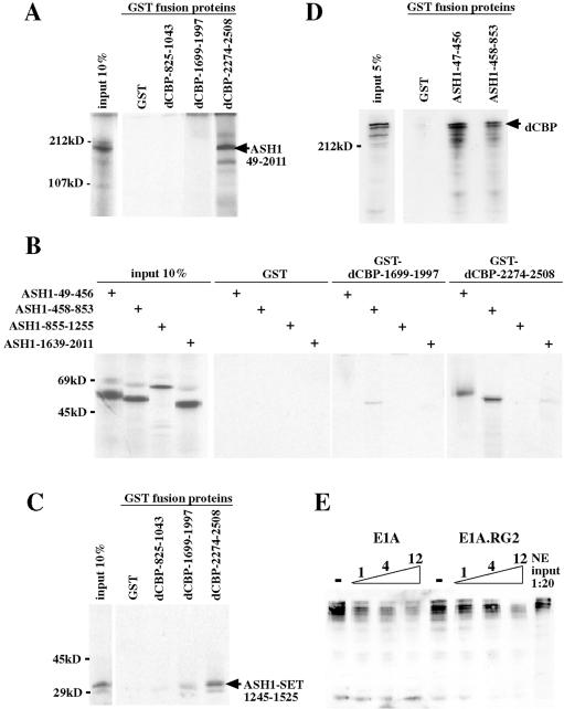
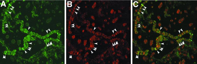
CREB-binding protein (CBP) is a coactivator for multiple transcription factors that transduce a variety of signaling pathways. Current models propose that CBP enhances gene expression by bridging the signal-responsive transcription factors with components of the basal transcriptional machinery and by augmenting the access of transcription factors to DNA through the acetylation of histones. To define the pathways and proteins that require CBP function in a living organism, we have begun a genetic analysis of CBP in flies. We have overproduced Drosophila melanogaster CBP (dCBP) in a variety of cell types and obtained distinct adult phenotypes. We used an uninflated-wing phenotype, caused by the overexpression of dCBP in specific central nervous system cells, to screen for suppressors of dCBP overactivity. Two genes with mutant versions that act as dominant suppressors of the wing phenotype were identified: the PKA-C1/DCO gene, encoding the catalytic subunit of cyclic AMP protein kinase, and ash1, a member of the trithorax group (trxG) of chromatin modifiers. Using immunocolocalization, we showed that the ASH1 protein is specifically expressed in the majority of the dCBP-overexpressing cells, suggesting that these proteins have the potential to interact biochemically. This model was confirmed by the findings that the proteins interact strongly in vitro and colocalize at specific sites on polytene chromosomes. The trxG proteins are thought to maintain gene expression during development by creating domains of open chromatin structure. Our results thus implicate a second class of chromatin-associated proteins in mediating dCBP function and imply that dCBP might be involved in the regulation of higher-order chromatin structure.


Figures

FIG. 1
FIG. 1
Uninflated-wing phenotype obtained by overexpressing dCBP in specific cells of the CNS using the GAL4-386 driver. (A) The uninflated-wing phenotype. (B to D) Expression of a LacZ reporter gene driven by GAL4-386 in larval tissues. β-Gal staining was detected in specific cells of the CNS (brain lobes and ventral ganglion) (B) but not in wing discs (C) or haltere/leg imaginal discs (D). (E to J) Analysis by laser confocal microscopy of dCBP wild-type expression and dCBP overexpression in the CNSs of third-instar larvae and pharate adult pupae. (E) dCBP ubiquitous expression in wild-type larval brain; (F) dCBP overexpression in specific cells in larval brain lobes and larval ventral ganglion; (G) overexpression in larval ventral ganglion at higher magnification; (H) overexpression in pupal brain including the optic lobe (OL); (I) overexpression in pupa thoracic ganglion with the prothorax (PT), the mesothorax (MS), the metathorax (MT), and the abdominal ganglion (AB) indicated; (J) overexpression in PT and MS at higher magnification. Magnifications, ×20 (E, F, H, and I) and ×40 (G and J).
FIG. 2
FIG. 2
dCBP overexpression with the GAL4-386 driver coincides with the expression of the neuropeptides FMRFamide and PHM. Arrows, colocalization of dCBP (rhodamine) and FMRFamide (fluorescein isothiocyanate [FITC]) in the larval (A) and pupal (C) ventral ganglion and colocalization of dCBP (rhodamine) and PHM (FITC) in the larval (B) and pupal (D) ventral ganglion.
FIG. 3
FIG. 3
Screen for deletions that can dominantly suppress the dCBP overexpression wing phenotype. Males carrying deficiencies for the second [Df(2)] and the third [Df(3)] chromosomes over a Balancer (B) were crossed to GAL4-386/MKRS; Tr21 (UAS-dCBP)/+ females with the uninflated-wing phenotype. Suppressed flies with the nonbalanced phenotype were analyzed. The Tr21 transgene on the fourth chromosome was not marked. To determine its presence in the suppressed flies, totally or partially suppressed males were crossed to w′ females to monitor the pale-orange eye color characteristic of flies with the Tr21 transgene. Examples of the mutant and suppressed-wing phenotypes are illustrated adjacent to the genotypes.
FIG. 4
FIG. 4
ASH1 expression coincides with dCBP overexpression in the CNS of GAL4-386/+; Tr21/+ larvae and pupae. An analysis by laser confocal microscopy was performed. (A to C) Colocalization in the thoracic region of a third-instar larval ventral ganglion. (A) ASH1 immunostaining with fluorescein isothiocyanate (FITC); (B) dCBP immunostaining with rhodamine; (C) merged image from panels A and B. (D to F) Colocalization in the thoracic CNSs, prothoraxes (PT), and mesothoraxes (MS) of pharate adult pupae. (D) ASH1-FITC immunostaining; (E) dCBP-rhodamine immunostaining; (F) merged image from panels D and E. (G to I) Colocalization in the thoracic CNSs metathoraxes (MT), and abdominal ganglia (AB) of pharate adult pupae. (G) ASH1-FITC immunostaining; (H) dCBP-rhodamine immunostaining; (I) merged image from panels G and H. Magnification, ×40.
FIG. 5
FIG. 5
CNS expression of ASH1 in dCBP-overexpressing or wild-type third-instar larvae, as analyzed by laser confocal microscopy. (A to C) Expression of ASH1 and dCBP in the larval ventral ganglion by dCBP-overexpressing cells. (A) ASH1 immunostaining with fluorescein isothiocyanate (FITC); (B) dCBP immunostaining with rhodamine; (C) merged image from panels A and B. (D to F) Expression of ASH1 and dCBP in the larva ventral ganglia of wild-type flies. (D) ASH1-FITC immunostaining; (E) dCBP-rhodamine immunostaining; (F) merged image from panels D and E. (G to I) Colocalization of ASH1 and dCBP in the nuclei of wild-type larval ganglion neurons. (G) ASH1-FITC immunostaining; (H) dCBP-rhodamine immunostaining; (I) merged image from panels G and H (arrowheads, fine haze of yellow colocalization). Magnifications, ×40 (A to F) and ×60 (G to I).
FIG. 6
FIG. 6
dCBP overexpression in a wild-type or ash122 heterozygous genetic background. Shown is an analysis by laser confocal microscopy of dCBP normal expression and overexpression in the ventral ganglia of third-instar larvae. (A) dCBP ubiquitous expression in wild-type larvae; (B) dCBP overexpression in GAL4-386/+; Tr21/+ larvae; (C) dCBP overexpression in GAL4-386/ash122; Tr21/+ larvae.
FIG. 7
FIG. 7
dCBP interacts with ASH1. (A) Equimolar amounts of immobilized GST and GST-dCBP fusion proteins were incubated with in vitro-translated 35S-labeled ASH1 protein (nearly full-length ASH1 protein; aa 49 to 2011). (B) Equimolar amounts of immobilized GST fusion proteins were incubated with various in vitro-translated 35S-labeled ASH1 fragments. The most C-terminal ASH1 fragment (aa 1639 to 2011) contains the PHD domain, but none of these fragments contain the SET domain. (C) Equimolar amounts of immobilized GST fusion proteins were incubated with the in vitro-translated 35S-labeled SET domain. (D) Equimolar amounts of immobilized GST and GST-ASH1 fusion proteins were incubated with Kc cell nuclear extracts. dCBP was detected by Western blotting using the dCBP chicken polyclonal antibody. (E) GST-ASH1-47-456 fusion protein was incubated with Kc cell nuclear extracts in the presence of 1, 4, or 12 μg of E1A or E1A-RG2. Similar results were obtained for GST-ASH1-458-853 (data not shown).
FIG. 8
FIG. 8
Endogenous dCBP and ASH1 proteins colocalize on wild-type polytene chromosomes. Shown is an analysis by laser confocal microscopy. (A) Chromosome arm showing localization of ASH1. (B) Chromosome arm from panel A showing localization of dCBP. (C) Merged image from panels A and B. Arrowheads, loci that colocalize both dCBP and ASH1. Magnification, ×60.

References

    1. Adamson A, Shearn A. Molecular genetic analysis of Drosophila ash2, a member of the trithorax group required for imaginal disc pattern formation. Genetics. 1996;144:621–633. - PMC - PubMed
    1. Akimaru H, Chen Y, Dai P, Hou D-X, Nonaka M, Smolik S M, Armstrong S, Goodman R H, Ishii S. Drosophila CBP is a co-activator of cubitus interruptus in hedgehog signalling. Nature. 1997;386:735–738. - PubMed
    1. Akimaru H, Hou D-X, Ishii S. Drosophila CBP is required for dorsal-dependent twist gene expression. Nat Genet. 1998;17:211–214. - PubMed
    1. Arany Z, Newsome D, Oldread E, Livingston D M, Eckner R. A family of transcriptional adaptor proteins targeted by the E1A oncoprotein. Nature. 1995;374:81–84. - PubMed
    1. Aza-Blanc P, Ramirez-Weber F-A, Laget M-P, Schwartz C, Kornberg T. Proteolysis that is inhibited by Hedgehog targets Cubitus interruptus protein to the nucleus and converts it to a repressor. Cell. 1997;89:1043–1053. - PubMed

Publication types

MeSH terms