. 2000 Dec;20(24):9364-75.
doi: 10.1128/MCB.20.24.9364-9375.2000.

Induced expression of Rnd3 is associated with transformation of polarized epithelial cells by the Raf-MEK-extracellular signal-regulated kinase pathway

Affiliations

Induced expression of Rnd3 is associated with transformation of polarized epithelial cells by the Raf-MEK-extracellular signal-regulated kinase pathway

S H Hansen et al. Mol Cell Biol. 2000 Dec.

Abstract

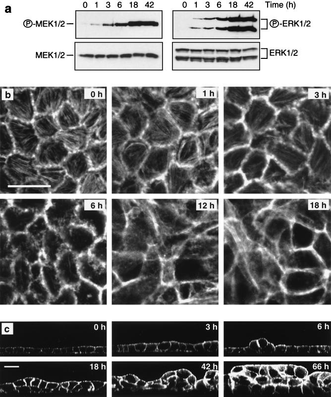
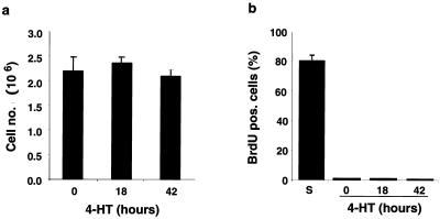
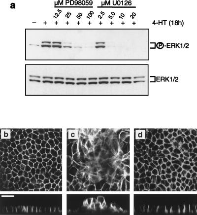
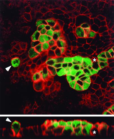
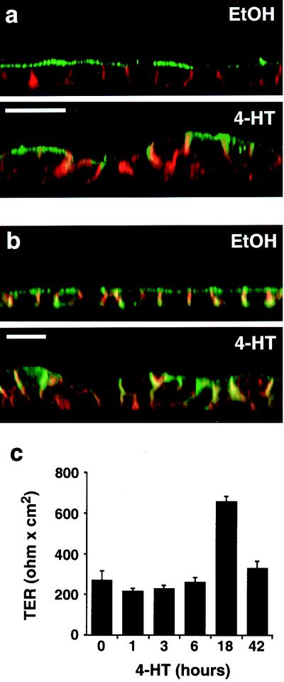
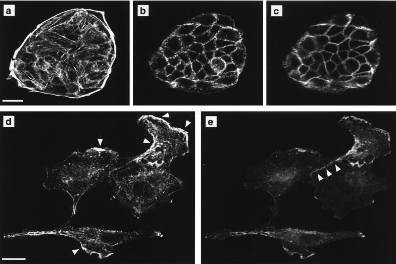
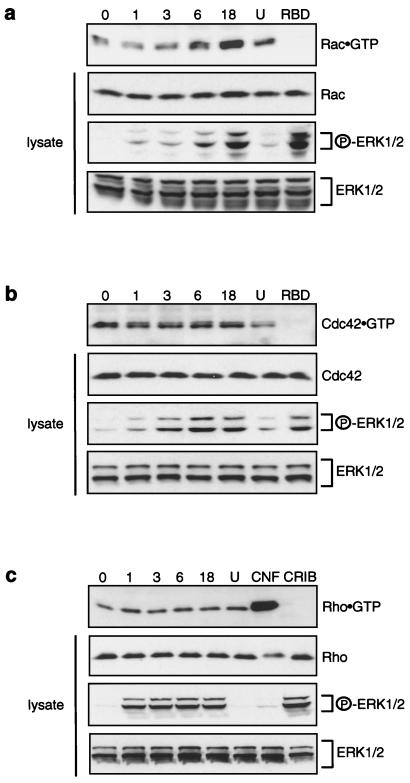
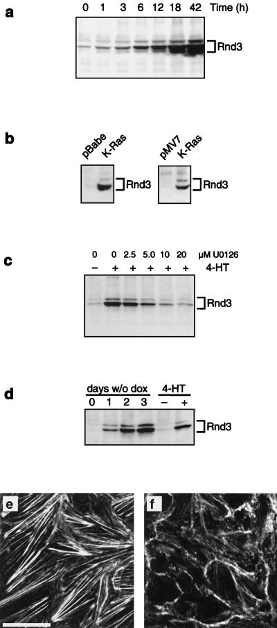
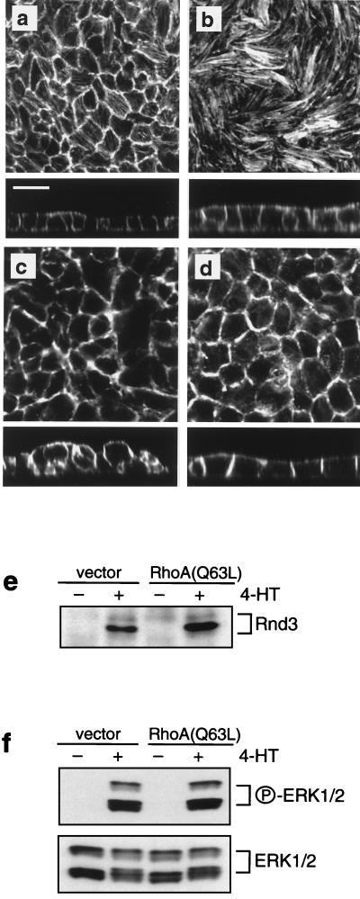
Madin-Darby canine kidney (MDCK) epithelial cells transformed by oncogenic Ras and Raf exhibit cell multilayering and alterations in the actin cytoskeleton. The changes in the actin cytoskeleton comprise a loss of actin stress fibers and enhanced cortical actin. Using MDCK cells expressing a conditionally active form of Raf, we have explored the molecular mechanisms that underlie these observations. Raf activation elicited a robust increase in Rac1 activity consistent with the observed increase in cortical actin. Loss of actin stress fibers is indicative of attenuated Rho function, but no change in Rho-GTP levels was detected following Raf activation. However, the loss of actin stress fibers in Raf-transformed cells was preceded by the induced expression of Rnd3, an endogenous inhibitor of Rho protein function. Expression of Rnd3 alone at levels equivalent to those observed following Raf transformation led to a substantial loss of actin stress fibers. Moreover, cells expressing activated RhoA failed to multilayer in response to Raf. Pharmacological inhibition of MEK activation prevented all of the biological and biochemical changes described above. Consequently, the data are consistent with a role for induced Rnd3 expression downstream of the Raf-MEK-extracellular signal-regulated kinase pathway in epithelial oncogenesis.


Figures

FIG. 1
FIG. 1
Time course of Raf-induced transformation of MDCK cells. Polarized MDCK cells expressing EGFPΔRaf-1:ER were treated with 1 μM 4-HT for the indicated periods of time. (a) Activation of MEK and ERK following Raf activation was assayed by Western blotting of whole cell lysates using activation-specific antibodies as described in Materials and Methods. (b and c) Confocal microscopy of MDCK cells labeled with fluorescent phalloidin revealed that the morphological alterations in response to Raf activation comprised progressive changes in cell shape (b and c), loss of actin stress fibers (b), increased cortical actin (b and c), and multilayering (c). (b) X-Y sections; scale bar, 20 μm. (c) X-Z sections; scale bar, 20 μm. All X-Y sections were sampled at the base of the cell monolayers.
FIG. 2
FIG. 2
Confluent MDCK cells are arrested both with and without Raf activation. (a) Cell counts evidenced that Raf activation in polarized MDCK cells does not result in a significant change in cell number. (b) BrdU incorporation to determine the fraction of cells in S phase carried out as described in Materials and Methods showed that polarized MDCK effectively remain arrested following activation of Raf. For comparison, BrdU incorporation in a subconfluent culture is shown (S).
FIG. 3
FIG. 3
MEK inhibitors abrogate transformation of MDCK cells by Raf. (a) MDCK cells expressing EGFPΔRaf-1:ER were either untreated or treated with 1 μM 4-HT for 18 h in the absence or presence of the indicated concentrations of the MEK inhibitor PD098059 or U0126. Activation of ERK was assayed by Western blotting of whole cell lysates using activation-specific antibodies as described in Materials and Methods. (b to d) EGFPΔRaf-1:ER-expressing MDCK cells were either untreated (b) or treated with 1 μM 4-HT for 18 h alone (c) or with 1 μM 4-HT for 18 h in the presence of 50 μM PD098059 (d). The cells were then fixed and labeled with fluorescent phalloidin and analyzed by confocal microscopy. All X-Y sections were sampled at the base of the cell monolayers. Scale bar, 20 μm.
FIG. 4
FIG. 4
Raf transformation of polarized MDCK cells is cell autonomous. MDCK cells expressing EGFPΔRaf-1:ER were mixed with the parental MDCK cell line at a ratio of 1:9; 42 h prior to experimentation, 1 μM 4-HT was added to the culture to activate Raf in the EGFPΔRaf-1:ER-expressing MDCK cells. With appropriate settings on the confocal microscope, EGFPΔRaf-1:ER-expressing MDCK cells can be identified based on the green fluorescence. In addition, the sample was labeled with fluorescent phalloidin to visualize the actin cytoskeleton (in red) in both parental and EGFPΔRaf-1:ER-expressing MDCK cells. The X-Y section was sampled close to the apical surface of the parental monolayer. Under these conditions, the EGFPΔRaf-1:ER-expressing MDCK cells invariably undergo morphological transformation and multilayer even when completely surrounded by parental cells (arrowhead). In contrast, the parental cells, except for changes in shape (asterisk) apparently inflicted by neighboring EGFPΔRaf-1:ER-expressing MDCK cells, show no signs of transformation and retain growth in monolayers. Scale bar, 20 μm.
FIG. 5
FIG. 5
Effects of Raf activation on cell polarity and on adherens and tight junctions. MDCK cells expressing EGFPΔRaf-1:ER were either untreated or treated with 1 μM 4-HT for 42 h. (a) The cells were fixed and labeled with fluorescent phalloidin to outline cell borders (in red) and immunolabeled to detect the 114-kDa apical marker (in green). Note the extensive redistribution of the 114-kDa apical marker to the lateral cell borders after Raf activation, where it colocalizes with cortical actin (in yellow-orange). Scale bar, 20 μm. (b) Cells were labeled with fluorescent phalloidin to outline cell borders (in green) and immunolabeled to detect the adherens junction constituent E-cadherin (in red). Note that despite the extensive cell-cell rearrangements occurring after Raf activation, E-cadherin still colocalizes with cortical actin at areas of cell-cell contact (in yellow-orange). Scale bar, 20 μm. (c) TER measurements in MDCK cells after activation of Raf for 0 to 42 h. As shown, the cells retain TER after Raf activation. The peak in TER observed after 18 h of Raf activation was observed in all six experiments carried out. Error bars indicate standard deviations (n = 3).
FIG. 6
FIG. 6
Raf transformation leads to spreading and scattering of MDCK cells. MDCK cells expressing EGFPΔRaf-1:ER grown at low cell density were either untreated (a to c) or treated with 1 μM 4-HT for 42 h (d and e). The cells were then fixed and labeled with fluorescent phalloidin to detect polymerized actin and immunolabeled to localize E-cadherin. (a and b) Phalloidin staining in optical sections through the basal (a) and apical (b) regions of untreated cells. (c) E-cadherin localization in the apical region of untreated cells. No E-cadherin labeling is detected in at the base of untreated cells (not shown). (d and e) Following activation of Raf, subconfluent MDCK cells become flattened, and both phalloidin (d) and E-cadherin (e) labeling is evident in a single optical section. Arrows in panel d indicate areas of apparent membrane ruffling. Note that E-cadherin colocalizes with polymerized actin at these areas. Arrowheads in panel e illustrate that E-cadherin is retained at areas of cell-cell contact in subconfluent Raf-transformed MDCK cells. Scale bars, 20 μm.
FIG. 7
FIG. 7
Effect of Raf activation on Rac1-, Cdc42-, and RhoA-GTP levels. Lanes represent GTP loading on endogenous Rac1 (a), Cdc42 (b), and RhoA (c) in MDCK cells following Raf activation for 0 to 18 h. Rac1-GTP and Cdc42-GTP levels were determined by the Pak3 CRIB assay (a and b, top panels). RhoA-GTP levels were assessed using the rhotekin RBD assay (c, top panel). Each experiment included one sample (lane U) where the cells were treated with 10 μM MEK inhibitor U0126 in addition to 1 μM 4-HT for 18 h. The rhotekin RBD assay (c) also included a sample (lane CNF) which was treated for 18 h with CNF1 only. Moreover, each experiment included one sample, in which Raf had been activated for 18 h, that was probed with the rhotekin RBD GST fusion protein for the CRIB assay (a and b, lanes RBD) and with the Pak3 CRIB GST fusion protein in the rhotekin RBD assay (c, lane CRIB). In addition, Western blots were carried out to detect Rac1, Cdc42, and RhoA in whole cell lysates, as well as phospho-ERK and total ERK (ERK1/2), and indicated on the right.
FIG. 8
FIG. 8
Induced expression of Rnd3 following Raf activation. (a) MDCK cells expressing EGFPΔRaf-1:ER were either untreated or treated with 1 μM 4-HT for the indicated periods of time, at which point the expression of Rnd3 was assessed by immunoprecipitation and Western blotting as described in Materials and Methods. (b) MDCK cells expressing K-Ras in either pBabe-Puro3 (pooled population) or pMV7 (clone) (63) were grown to confluency, and expression of Rnd3 was assessed by immunoprecipitation and Western blotting. (c) MDCK cells expressing EGFPΔRaf-1:ER were either untreated or treated with 1 μM 4-HT for 18 h in the absence or presence of the indicated concentrations of the MEK inhibitor U0126. The expression of Rnd3 was assessed by immunoprecipitation and Western blotting. (d) MDCK cells expressing Rnd3 under the control of the Tet-off system were cultured in the presence (0) or absence of doxycycline for 1 to 3 days, at which time the expression of Rnd3 was assessed by immunoprecipitation and Western blotting. As a control, the expression of Rnd3 was measured in extracts of MDCK cells expressing EGFPΔRaf-1:ER that were either untreated or treated with 1 μM 4-HT for 42 h. (e and f) MDCK cells expressing Rnd3 under the control of the Tet-off system were cultured in the presence (e) or absence (f) of doxycycline for 2 days. At this time, the cells were labeled with fluorescent phalloidin to detect polymerized actin and analyzed by confocal microscopy. Both optical sections were sampled at the base of the cell monolayers. Scale bar, 20 μm.
FIG. 9
FIG. 9
Constitutive expression of RhoA(Q63L) prevents Raf-induced multilayering. (a to d) MDCK cells expressing EGFPΔRaf-1:ER were transduced with either empty vector, pWZLNeo (a and c), or the same vector encoding RhoA(Q63L) (b and d), and clonal cell lines were derived. The cells were then either untreated (a and b) or treated with 1 μM 4-HT for 42 h (c and d), at which time the cells were labeled with fluorescent phalloidin and analyzed by confocal microscopy. (e and f) The cells described above were either untreated or treated with 1 μM 4-HT for 42 h, at which time the expression of Rnd3 (e) and activation of ERK1 and -2 (f) were assessed as described in Materials and Methods. All X-Y sections were sampled at the base of the cell monolayers. Scale bar, 20 μm.

Similar articles

Cited by

References

    1. Alessi D R, Cuenda A, Cohen P, Dudley D T, Saltiel A R. PD 098059 is a specific inhibitor of the activation of mitogen-activated protein kinase kinase in vitro and in vivo. J Biol Chem. 1995;270:27489–27894. - PubMed
    1. Amano M, Chihara K, Kimura K, Fukata Y, Nakamura N, Matsuura Y, Kaibuchi K. Formation of actin stress fibers and focal adhesions enhanced by Rho-kinase. Science. 1997;275:1308–1311. - PubMed
    1. Bagrodia S, Taylor S J, Jordon K A, Van Aelst L, Cerione R A. A novel regulator of p21-activated kinases. J Biol Chem. 1998;273:23633–23636. - PubMed
    1. Balcarova-Ständer J, Pfeiffer S E, Fuller S D, Simons K. Development of cell surface polarity in the epithelial Madin-Darby canine kidney (MDCK) cell line. EMBO J. 1984;3:2687–2694. - PMC - PubMed
    1. Balkovetz D F, Pollack A L, Mostov K E. Hepatocyte growth factor alters the polarity of Madin-Darby canine kidney cell monolayers. J Biol Chem. 1997;272:3471–3477. - PubMed

Publication types

MeSH terms