. 2000 Dec;20(24):9391-8.
doi: 10.1128/MCB.20.24.9391-9398.2000.

Multiple lysine mutations in the C-terminal domain of p53 interfere with MDM2-dependent protein degradation and ubiquitination

Affiliations

Multiple lysine mutations in the C-terminal domain of p53 interfere with MDM2-dependent protein degradation and ubiquitination

S Nakamura et al. Mol Cell Biol. 2000 Dec.

Abstract

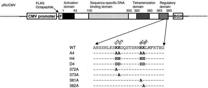
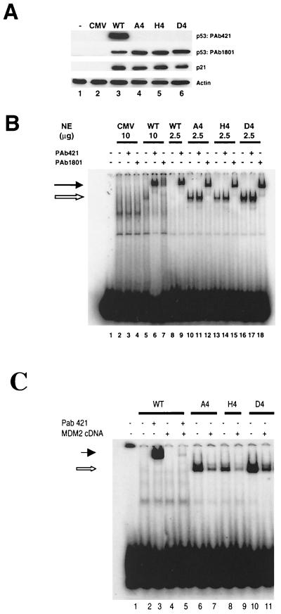
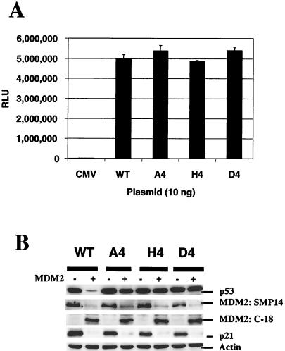
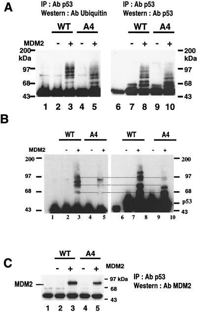
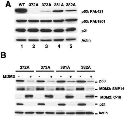
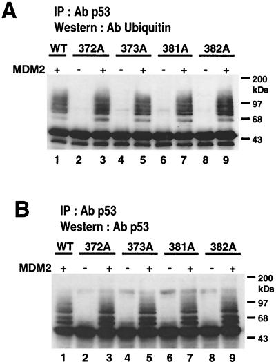
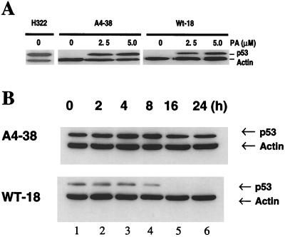
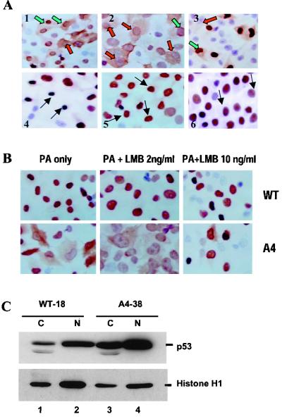
To investigate the effect of mutations in the p53 C-terminal domain on MDM2-mediated degradation, we introduced single and multiple point mutations into a human p53 cDNA at four putative acetylation sites (amino acid residues 372, 373, 381, and 382). Substitution of all four lysine residues by alanines (the A4 mutant) and single lysine-to-alanine substitutions were functional in sequence-specific DNA binding and transactivation; however, the A4 mutant protein was resistant to MDM2-mediated degradation, whereas the single lysine substitutions were not. Although the A4 mutant protein and the single lysine substitutions both bound MDM2 reasonably well, the single lysine substitutions underwent normal MDM2-dependent ubiquitination, whereas the A4 protein was inefficiently ubiquitinated. In addition, the A4 mutant protein was found in the cytoplasm as well as in the nucleus of a subpopulation of cells, unlike wild-type p53, which is mostly nuclear. The partially cytoplasmic distribution of A4 mutant protein was not due to a defect in nuclear import because inhibition of nuclear export by leptomycin B resulted in nuclear accumulation of the protein. Taken together, the data suggest that mutations in the putative acetylation sites of the p53 C-terminal domain interfere with ubiquitination, thereby regulating p53 degradation.


Figures

FIG. 1
FIG. 1
Structure of an expression plasmid carrying wild-type p53 cDNA. Functional domains of human p53 and positions of mutations (highlighted) are displayed. BGH, BGH poly(A) signal; WT, wild-type p53.
FIG. 2
FIG. 2
Expression and DNA-binding activity of wild-type and mutant p53 proteins. (A) Western blot analysis of p53 and p21 protein expression in H1299 cells transiently transfected with wild-type or mutated p53 cDNAs as indicated. At 24 h after transfection, cell extracts were prepared, resolved by SDS–10% PAGE, and transferred to a nitrocellulose membrane. The membrane was probed with the indicated antibodies. (B) Nuclear extracts were prepared from H1299 cells transfected with the indicated plasmids and used for EMSA. The 32P-labeled p53CON oligonucleotide was incubated in the absence (lane 1) or presence of the indicated nuclear extracts (lanes 2 to 18). Incubations were performed either in the absence of PAb421 and PAb1801 (lanes 1, 2, 5, 8, 10, 13, and 16) or in the presence of either PAb421 (lanes 3, 6, 9, 11, 14, and 17) or PAb1801 (lanes 4, 7, 12, 15, and 18). (C) Overexpression of exogenous mouse MDM2 inhibited p53 DNA binding activity. Mutants also showed inhibition of their DNA binding activity (lanes 7, 9, and 11). The mixtures were resolved by PAGE and visualized using autoradiography. The open and closed arrows indicate the positions of the shifted and supershifted complexes, respectively.
FIG. 3
FIG. 3
Transactivation and stability of mutant p53 in the presence of MDM2 overexpression. (A) H1299 cells were transiently transfected with 10 ng of each p53 construct and 1 μg of pGUP.PA.8-p53CON plasmid DNA. After 24 h, cells were lysed and assayed for luciferase activity. The values presented here are representative data from triplicate experiments. Assays were performed twice independently and showed similar results. (B) H1299 cells were transfected with 0.5 μg of p53 plasmid and 4.5 μg of pRc/CMV empty vector or MDM2 cDNA plasmid. At 24 h after transfection, cell extracts were prepared, and expression of p53, expression of MDM2, and induction of p21 were analyzed by Western blot analysis using PAb1801, MDM2 mouse monoclonal SMP14, and rabbit polyclonal antibody C-18 (Santa Cruz), and p21 monoclonal antibody, respectively.
FIG. 4
FIG. 4
MDM2-mediated ubiquitination of wild-type p53 and A4 mutant. (A) H1299 cells were transfected with 5 μg of MDM2 plasmid and 5 μg of either wild-type p53- or A4 mutant-expressing plasmid. At 24 h after transfection, cell extracts were prepared as described in Materials and Methods and then immunoprecipitated with DO-1 anti-p53 antibody. The immunoprecipitates were subjected to Western blot analysis. A blot was probed first with ubiquitin monoclonal antibody P1A6 (lanes 1 to 5), then stripped, and then reprobed with Bp53-12 (lanes 6 to 10). Lanes 1 and 6, immunoprecipitates from untransfected cells serving as negative controls and showing the position of the IgG heavy chain immediately below the p53 protein band. (B) To separate the p53-ubiquitinated bands more clearly, a longer gel was run using the same strategy as above. Ubiquitin p53 complexes were marked. (C) A blot was probed first with anti-MDM2 antibody (upper), then stripped, and then reprobed with anti-p53 antibody (lower panel). Lane 1, immunoprecipitates from untransfected cells serving as negative controls.
FIG. 5
FIG. 5
Expression of single-lysine mutants and their degradation in the presence of MDM2 overexpression. H1299 cells were transfected with either 5 μg of each indicated plasmid or a combination of plasmids as indicated. At 24 h after transfection, cell extracts were prepared, and expression of p53, expression of MDM2, and induction of p21 were analyzed by Western blot analysis. MDM2 protein was detected by mouse monoclonal antibody SMP14 and rabbit polyclonal antibody C-18 (Santa Cruz). WAF1 monoclonal antibody EA10 was used for detecting p21. (A) Expression of wild-type p53 and single-lysine mutants. (B) Single-lysine mutant protein expression in the presence or absence of MDM2.
FIG. 6
FIG. 6
Efficient ubiquitination of single-lysine mutants and their degradation by MDM2. H1299 cells were transfected with 5 μg of MDM2 plasmid and 5 μg of plasmid expressing either wild-type p53 or a single-lysine mutant. At 24 h after transfection, cell extracts were prepared and immunoprecipitated with DO-1 anti-p53 monoclonal antibody as described above. The immunoprecipitates were subjected to Western blot analysis. (A) Blot probed with ubiquitin monoclonal antibody P1A6. (B) Same blot stripped and reprobed with anti-p53 antibody.
FIG. 7
FIG. 7
PA-induced expression of p53 protein in H1299 cells stably transfected with WT-18 and A4-38 as shown by immunoblot analysis of cell lysates. (A) p53 protein in WT-18 and A4-38 cells incubated for 24 h in the presence of as indicated PA. Actin protein served as a loading control. An H322 lung cancer cell line carrying a spontaneously mutated p53 gene was used as a positive control. (B) The stability of wild-type and A4 p53 protein in these clones was examined. The p53 protein in these cells was induced by 5 μM PA for 24 h. Induced cells were then incubated with 10 μg of cycloheximide per ml for different time intervals and then processed to prepare total cell extracts. p53 and actin proteins were detected by Western blot analysis using monoclonal antibodies as described earlier. (Upper panel) A4-38 cells. (Lower panel) WT-18 cells. Lanes 1 to 6 show p53 and actin proteins after 0, 2, 4, 8, 16, and 24 h of cycloheximide treatment, respectively.
FIG. 8
FIG. 8
View of immunohistochemically stained wild-type and mutant p53 protein in PA-induced cells. (A) Cells were grown on a culture slide and treated with 5 μM PA for 24 h. They were then stained with PAb1801 anti-p53 antibody. In the majority of cells, mutant p53 protein was distributed in both the nucleus and the cytoplasm of A4-38 clones (panels 1 and 2), while wild-type p53 was predominantly confined to the nucleus despite intense nuclear accumulation (panels 4 and 5). A4-41, another clone of A4, showed a similar cytoplasmic distribution (panel 5). A lung cancer cell line with a spontaneous mutation at codon 248 was used as a positive control; in this line, mutant p53 was restricted to the nucleus (panel 6). The red-filled arrow shows a strong cytoplasmic distribution of A4 mutant protein and a much lower nuclear accumulation. The green-filled arrow indicates the strong nuclear accumulation of p53 but cytoplasmic distribution. The black-filled arrow indicates strong nuclear localization of p53 with no cytoplasmic distribution. (B) Nuclear localization of leptomycin B-induced p53 protein. WT-18 cells (a, b, and c) and A4-38 cells (d, e, and f) were left untreated (a and d) or were treated with 2 ng of leptomycin B (b and e) or 10 ng of leptomycin B per ml for 18 h prior to fixation. p53 protein was detected in cells using anti-p53 monoclonal antibody as primary antibody and goat anti-mouse antibody as secondary antibody. Cells were also processed for immunohistochemical staining using an ABC kit. In both cases, the induced p53 protein was localized to the nucleus of the cells following leptomycin B treatment. (C) WT-18 and A4-38 cells were induced with PA for 24 h and fractionated into cytosolic (C) and nuclear (N) extracts as described in Materials and Methods. The extracts from an equivalent number of cells for each cell line were loaded onto a SDS-polyacrylamide gel and transferred to a nitrocellulose membrane. The membrane was then probed with anti-p53 antibody and reprobed with histone HI monoclonal antibody.

Similar articles

Cited by

References

    1. Ciechanover A. The ubiquitin-proteasome pathway: on protein death and cell life. EMBO J. 1998;17:7151–7160. - PMC - PubMed
    1. El Deiry W S, Tokino T, Velculescu V E, Levy D B, Parsons R, Trent J M, Lin D, Mercer W E, Kinzler K W, Vogelstein B. WAF1, a potential mediator of p53 tumor suppression. Cell. 1993;75:817–825. - PubMed
    1. Freedman D A, Levine A J. Nuclear export is required for degradation of endogenous p53 by MDM2 and human papillomavirus E6. Mol Cell Biol. 1998;18:7288–7293. - PMC - PubMed
    1. Funk W D, Pak D T, Karas R H, Wright W E, Shay J W. A transcriptionally active DNA-binding site for human p53 protein complexes. Mol Cell Biol. 1992;12:2866–2871. - PMC - PubMed
    1. Gu W, Roeder R G. Activation of p53 sequence-specific DNA binding by acetylation of the p53 C-terminal domain. Cell. 1997;90:595–606. - PubMed

Publication types

MeSH terms

LinkOut - more resources