

Intact MutY and its catalytic domain differentially contact with A/8-oxoG-containing DNA
- PMID: 11095667
- PMCID: PMC115170
- DOI: 10.1093/nar/28.23.4593
Intact MutY and its catalytic domain differentially contact with A/8-oxoG-containing DNA
Abstract
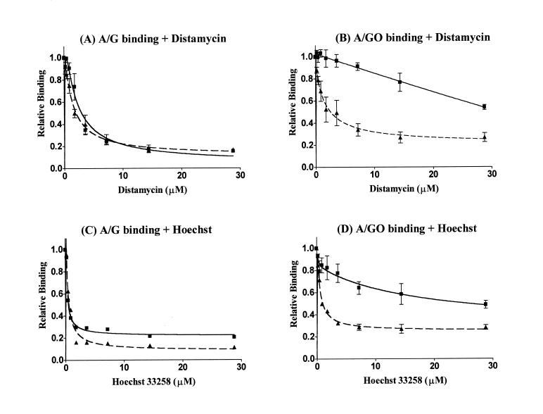
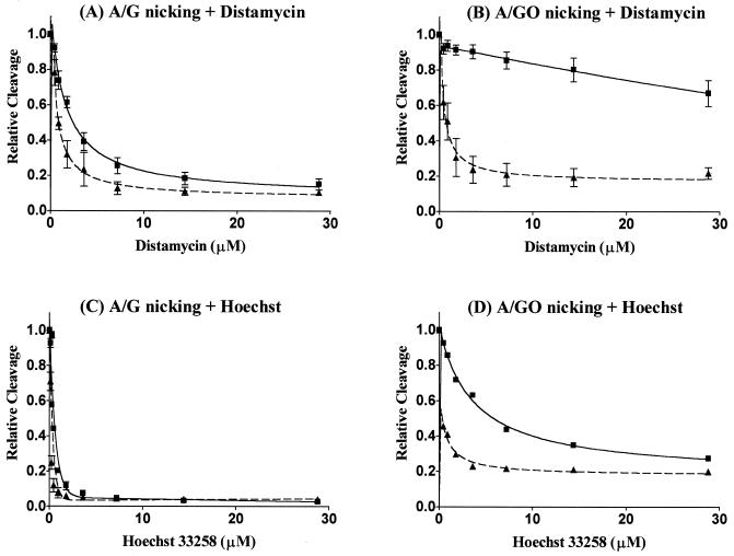
Escherichia coli MutY is an adenine and a weak guanine DNA glycosylase active on DNA substrates containing A/G, A/8-oxoG, A/C or G/8-oxoG mismatches. A truncated form of MutY (M25, residues 1-226) retains catalytic activity; however, the C-terminal domain of MutY is required for specific binding to the 8-oxoG and is critical for mutation avoidance of oxidative damage. Using alkylation interference experiments, the determinants of the truncated and intact MutY were compared on A/8-oxoG-containing DNA. Several purines within the proximity of mismatched A/8-oxoG show differential contact by the truncated and intact MutY. Most importantly, methylation at the N7 position of the mismatched 8-oxoG and the N3 position of mismatched A interfere with intact MutY but not with M25 binding. The electrostatic contacts of MutY and M25 with the A/8-oxoG-containing DNA substrates are drastically different as shown by ethylation interference experiments. Five consecutive phosphate groups surrounding the 8-oxoG (one on the 3' side and four on the 5' side) interact with MutY but not with M25. The activities of the truncated and intact MutY are modulated differently by two minor groove-binding drugs, distamycin A and Hoechst 33258. Both distamycin A and Hoechst 33258 can inhibit, to a similar extent, the binding and glycosylase activities of MutY and M25 on A/G mismatch. However, binding and glycosylase activities on A/8-oxoG mismatch of intact MutY are inhibited to a lesser degree than those of M25. Overall, these results suggest that the C-terminal domain of MutY specifies additional contact sites on A/GO-containing DNA that are not found in MutY-A/G and M25-A/8-oxoG interactions.
Figures


Similar articles
-
Specific recognition of A/G and A/7,8-dihydro-8-oxoguanine (8-oxoG) mismatches by Escherichia coli MutY: removal of the C-terminal domain preferentially affects A/8-oxoG recognition.Biochemistry. 1996 Dec 24;35(51):16665-71. doi: 10.1021/bi960843w. Biochemistry. 1996. PMID: 8988002
-
The C-terminal domain of MutY glycosylase determines the 7,8-dihydro-8-oxo-guanine specificity and is crucial for mutation avoidance.J Biol Chem. 2000 Mar 24;275(12):8448-55. doi: 10.1074/jbc.275.12.8448. J Biol Chem. 2000. PMID: 10722679
-
The C-terminal domain of Escherichia coli MutY is involved in DNA binding and glycosylase activities.Nucleic Acids Res. 2003 Jun 15;31(12):3038-49. doi: 10.1093/nar/gkg434. Nucleic Acids Res. 2003. PMID: 12799430 Free PMC article.
-
Potential double-flipping mechanism by E. coli MutY.Prog Nucleic Acid Res Mol Biol. 2001;68:349-64. doi: 10.1016/s0079-6603(01)68111-x. Prog Nucleic Acid Res Mol Biol. 2001. PMID: 11554310 Review.
-
Repair of 8-oxo-7,8-dihydroguanine in prokaryotic and eukaryotic cells: Properties and biological roles of the Fpg and OGG1 DNA N-glycosylases.Free Radic Biol Med. 2017 Jun;107:179-201. doi: 10.1016/j.freeradbiomed.2016.11.042. Epub 2016 Nov 27. Free Radic Biol Med. 2017. PMID: 27903453 Review.
Cited by
-
Physical and functional interactions between Escherichia coli MutY and endonuclease VIII.Biochem J. 2006 Jan 1;393(Pt 1):381-7. doi: 10.1042/BJ20051133. Biochem J. 2006. PMID: 16201966 Free PMC article.
-
MUTYH DNA glycosylase: the rationale for removing undamaged bases from the DNA.Front Genet. 2013 Feb 28;4:18. doi: 10.3389/fgene.2013.00018. eCollection 2013. Front Genet. 2013. PMID: 23450852 Free PMC article.
-
Inhibitors of DNA Glycosylases as Prospective Drugs.Int J Mol Sci. 2020 Apr 28;21(9):3118. doi: 10.3390/ijms21093118. Int J Mol Sci. 2020. PMID: 32354123 Free PMC article. Review.
-
Direct Measurement of 8OG Syn-Anti Flips in Mutagenic 8OG·A and Long-Range Damage-Dependent Hoogsteen Breathing Dynamics Using 1H CEST NMR.J Phys Chem B. 2024 May 2;128(17):4087-4096. doi: 10.1021/acs.jpcb.4c00316. Epub 2024 Apr 22. J Phys Chem B. 2024. PMID: 38644782 Free PMC article.
-
Direct Measurement of 8OG syn-anti Flips in Mutagenic 8OG•A and Long-Range Damage-Dependent Hoogsteen Breathing Dynamics Using 1H CEST NMR.bioRxiv [Preprint]. 2024 Jan 16:2024.01.15.575532. doi: 10.1101/2024.01.15.575532. bioRxiv. 2024. Update in: J Phys Chem B. 2024 May 2;128(17):4087-4096. doi: 10.1021/acs.jpcb.4c00316. PMID: 38293035 Free PMC article. Updated. Preprint.
References
-
- Tchou J. and Grollman,A.P. (1993) Repair of DNA containing the oxidatively-damaged base 8-hydroxyguanine. Mutat. Res., 299, 277–287. - PubMed
-
- Li X., Wright,P.M. and Lu,A.-L. (2000) The C-terminal domain of MutY glycosylase determines the 7,8-dihydro-8-oxo-guanine specificity and is crucial for mutation avoidance. J. Biol. Chem., 275, 8448–8455. - PubMed
Publication types
MeSH terms
Substances
Grants and funding
LinkOut - more resources
Full Text Sources
