Comparative Study
. 2000 Dec 1;28(23):4698-708.
doi: 10.1093/nar/28.23.4698.

Comparative analysis of more than 3000 sequences reveals the existence of two pseudoknots in area V4 of eukaryotic small subunit ribosomal RNA

Affiliations
Comparative Study

Comparative analysis of more than 3000 sequences reveals the existence of two pseudoknots in area V4 of eukaryotic small subunit ribosomal RNA

J Wuyts et al. Nucleic Acids Res. .

Abstract

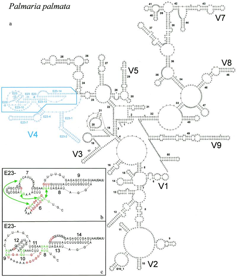
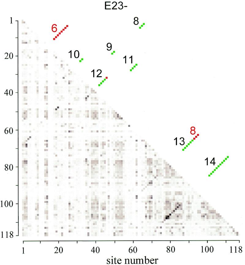
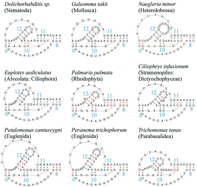
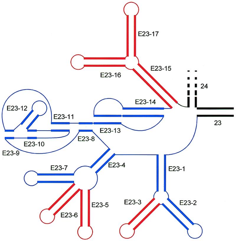
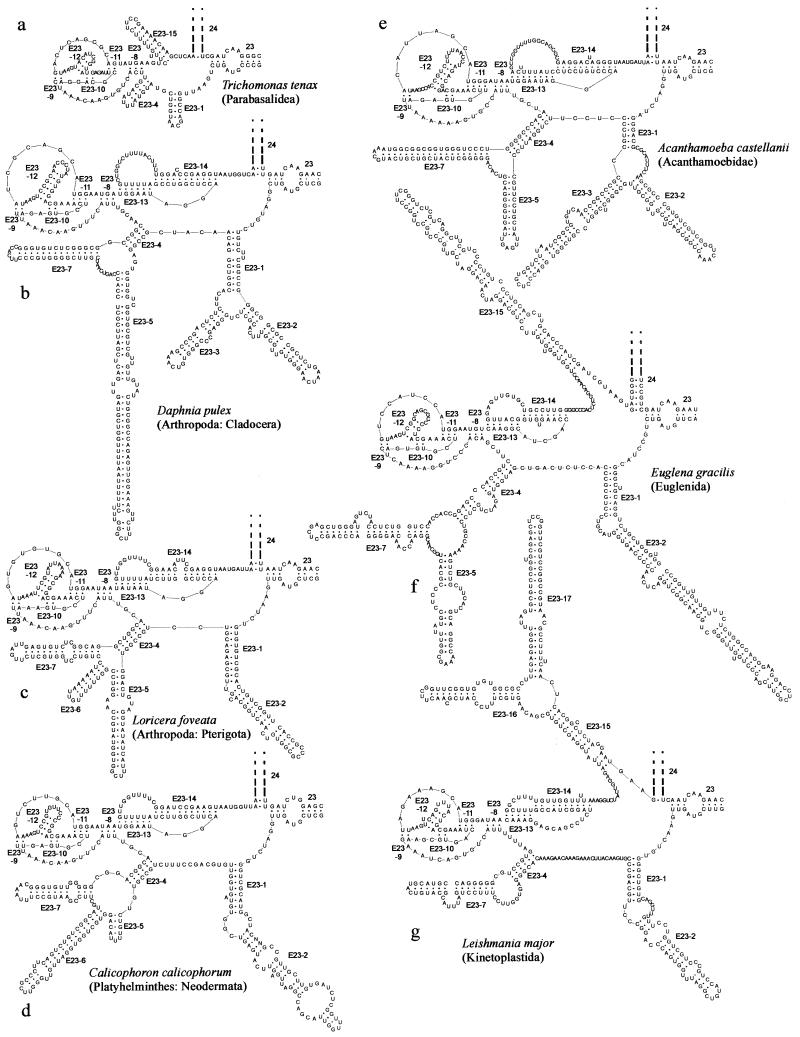
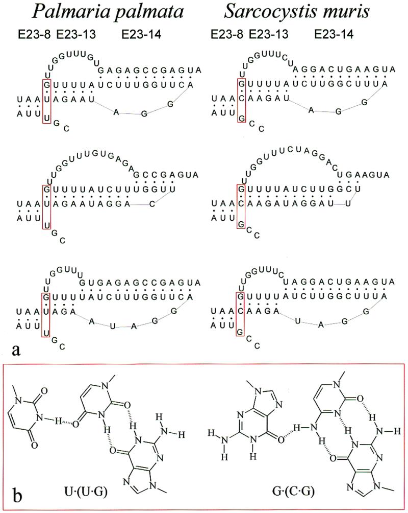
The secondary structure of V4, the largest variable area of eukaryotic small subunit ribosomal RNA, was re-examined by comparative analysis of 3253 nucleotide sequences distributed over the animal, plant and fungal kingdoms and a diverse set of protist taxa. An extensive search for compensating base pair substitutions and for base covariation revealed that in most eukaryotes the secondary structure of the area consists of 11 helices and includes two pseudoknots. In one of the pseudoknots, exchange of base pairs between the two stems seems to occur, and covariation analysis points to the presence of a base triple. The area also contains three potential insertion points where additional hairpins or branched structures are present in a number of taxa scattered throughout the eukaryotic domain.


Figures

Figure 1
Figure 1
New secondary structure model illustrated with P.palmata SSU rRNA. (a) Complete model with indication of variable areas V1–V9. V6 is variable only in prokaryotic SSU rRNA where helix 37 is branched. Area V4 is in blue; base pair compensation and base covariation was examined systematically for the boxed part, details of which are shown in the inserts (b) and (c). (b) Old model and helix numbering for part of area V4. Abolished base pairs are in red and new ones, connected by arrows, are in green. (c) New model and helix numbering for part of area V4. Color conventions as in (b). The numbering system was changed to fit the new helix succession and to allow numbering of extra helices present in certain taxa (see Table 3). The correspondence between old and new helix number is as follows: helix E23-6 of the old model is dismantled; E23-7 (old) is transformed into helices E23-11 and 12 (new); E23-8 (old) is partly conserved as E23-13 (new); E23-9 (old) becomes E23-14 (new). Helices E23-8,9 and 10 of the new model do not exist in the old model.
Figure 2
Figure 2
Matrix of base pair compensation and covariation strength. The lower half matrix consists of dots with a darkness proportional to the value of √ϕC measured for each pair that can be formed by the 118 sites lying between helix E23-4 and helix 24 and occcupied by nucleotides in at least 90% of the 3253 sequences examined. There are 256 shades of gray ranging from white (value 0) to black (the highest value measured, 0.8016). Most helices showing base pair compensation are visible as diagonals of darker dots. The upper half matrix, symmetrical with the lower half, shows the position of the helices E23-8 to 14 of the new secondary structure model (Fig. 1c) as green dots. The red dots are abolished base pairs with old helix numbers (Fig. 1b).
Figure 3
Figure 3
Base pair compensation in helices E23-8 to E23-12. Helices E23-8 to E23-12 comprise 15 base pairs in most species. Fourteen of these, in red in the P.palmata structure in the centre, show compensation in other species, as can be seen in the eight structures surrounding it. The first base pair of helix E23-11 (U·A, at right in the figure), though subject to compensation, is non-complementary in ∼1/4 of the sequences. In Trichonomas tenax this base pair cannot be formed if it is assumed that the pseudoknot loop between helices E23-10 and 11 must contain at least one nucleotide (G in this case). Compensation of the second base pair of helix E23-11 occurs between the structures of Ciliophrys infusionum (C·G) and Peranema trichophorum (U·A). The last base pair (A·U) is not compensated in presently known species but becomes G·U in two species. See also Table 2.
Figure 4
Figure 4
Position of exceptional helices in area V4. Double stranded areas are drawn as thick parallel lines, single stranded areas as thin lines. Universal helices 23 and 24 are drawn in black, eukaryote-specific helices are in color, blue for those present in the majority of species, red for those present only in specific taxa (cf. Table 3).
Figure 5
Figure 5
Examples of exceptional structures in area V4 in specific taxa. (a) Parabasalidea; (b) Cladocera; (c) Pterigota; (d) Neodermata; (e) Acanthamoebidae; (f) Euglenida; (g) Kinetoplastida. Cf. Table 3.
Figure 6
Figure 6
Possible dynamic structure and base triple in pseudoknot E23-13-14. (a) The structure of helix E23-8 and of the pseudoknot E23-13-14 is shown for two species, the red alga P.palmata and the apicomplexan Sarcocystis muris. The uppermost structure corresponds to the model of Figure 1a and c. In the middle structures the boundary between the pseudoknot helices is shifted to the right, in the lower structure to the left, by disrupting base pairs of one helix in favour of the other. Postulated base triples U·(U·G) in P.palmata and G·(C·G) in S.muris are boxed in red. (b) Structural formulas of the most isomorphic forms of the base triples U·(U·G) on the left and G·(C·G) on the right.

References

    1. Gutell R.R., Weiser,B., Woese,C.R. and Noller,H.F. (1985) Progr. Nucleic Acids Res. Mol. Biol., 32, 155–216. - PubMed
    1. De Rijk P., Neefs,J.M., Van de Peer,Y. and De Wachter,R. (1992) Nucleic Acids Res., 20, Suppl., 2075–2089. - PMC - PubMed
    1. Gutell R.R., Gray,M.W. and Schnare,M.N. (1993) Nucleic Acids Res., 21, 3055–3074. - PMC - PubMed
    1. De Rijk P., Van de Peer,Y., Chapelle,S. and De Wachter,R. (1994) Nucleic Acids Res., 22, 3495–3501. - PMC - PubMed
    1. Gutell R.R., Larsen,N. and Woese,C.R. (1994) Microbiol. Rev., 58, 10–26. - PMC - PubMed

Publication types