. 2000 Dec 1;28(23):4717-24.
doi: 10.1093/nar/28.23.4717.

Error-prone lesion bypass by human DNA polymerase eta

Affiliations

Error-prone lesion bypass by human DNA polymerase eta

Y Zhang et al. Nucleic Acids Res. .

Abstract

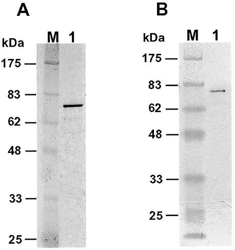
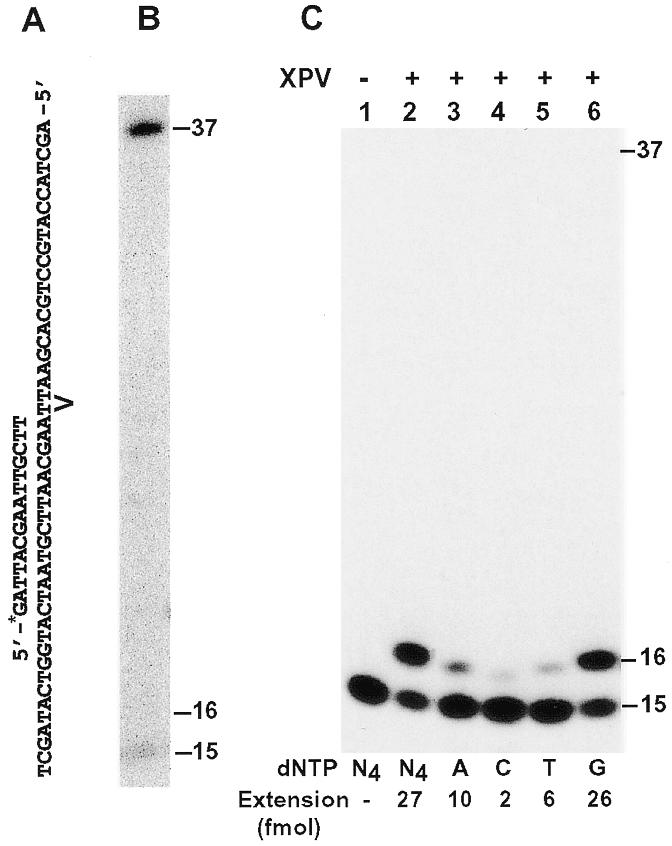
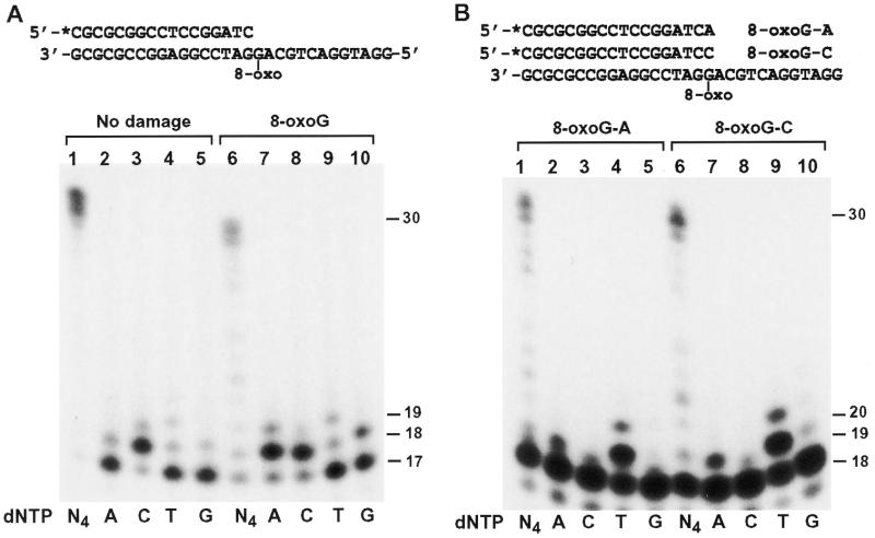
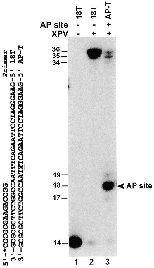
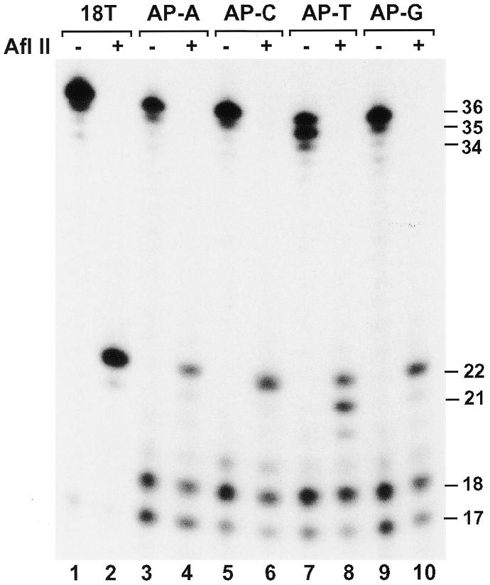
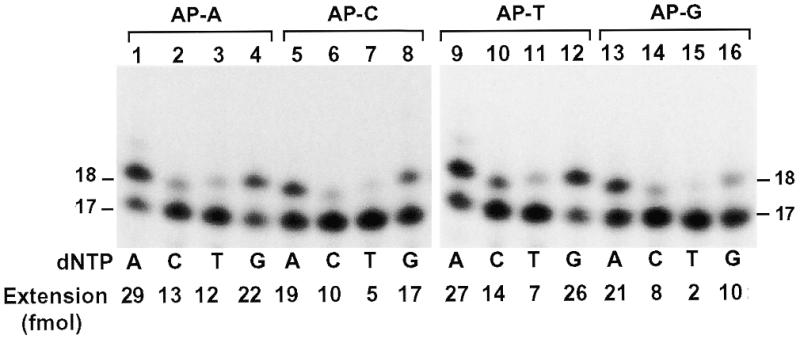
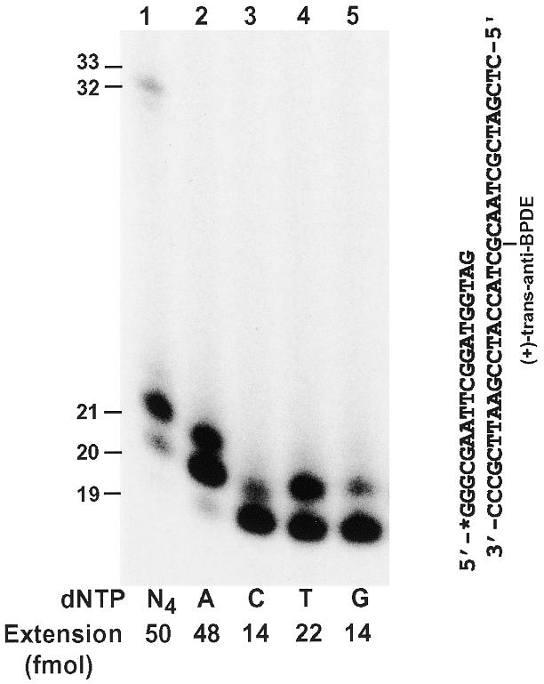
DNA lesion bypass is an important cellular response to genomic damage during replication. Human DNA polymerase eta (Pol(eta)), encoded by the Xeroderma pigmentosum variant (XPV) gene, is known for its activity of error-free translesion synthesis opposite a TT cis-syn cyclobutane dimer. Using purified human Pol(eta), we have examined bypass activities of this polymerase opposite several other DNA lesions. Human Pol(eta) efficiently bypassed a template 8-oxoguanine, incorporating an A or a C opposite the lesion with similar efficiencies. Human Pol(eta) effectively bypassed a template abasic site, incorporating an A and less frequently a G opposite the lesion. Significant -1 deletion was also observed when the template base 5' to the abasic site is a T. Human Pol(eta) partially bypassed a template (+)-trans-anti-benzo[a]pyrene-N:(2)-dG and predominantly incorporated an A, less frequently a T, and least frequently a G or a C opposite the lesion. This specificity of nucleotide incorporation correlates well with the known mutation spectrum of (+)-trans-anti-benzo[a]pyrene-N:(2)-dG lesion in mammalian cells. These results show that human Pol(eta) is capable of error-prone translesion DNA syntheses in vitro and suggest that Pol(eta) may bypass certain lesions with a mutagenic consequence in humans.


Figures

Figure 1
Figure 1
Analyses of purified human Polη. (A) Purified human Polη (80 ng) was analyzed by electrophoresis on a 10% SDS–polyacrylamide gel and visualized by silver staining. Protein size markers (lane M) are indicated on the left. (B) Purified human Polη (16 ng) was analyzed by a western blot using a mouse monoclonal antibody against the His6 tag. Protein size markers (lane M) are indicated on the left.
Figure 2
Figure 2
Response of human Polη to a TT dimer and a TT (6-4) photoproduct in template DNA. (A) Primed DNA template containing a TT dimer or a TT (6-4) photoproduct as indicated at the TT sequence. The 15mer primer was labeled with 32P at its 5′-end (*). (B) A lesion bypass assay was performed on the TT dimer template with 0.8 ng of purified human Polη. (C) DNA polymerase assays were performed with 0.8 ng (10 fmol, 1 nM) human Polη using the template (50 fmol) containing a TT (6-4) photoproduct. Polymerase reactions were carried out in the presence of a single deoxyribonucleoside triphosphate dATP (lane 3), dCTP (lane 4), dTTP (lane 5), dGTP (lane 6) or all four dNTPs (lane 2). Lane 1, control reaction without DNA polymerase. Quantitation of extended primers is shown at the bottom of the gel. DNA size markers in nucleotides are indicated on the right.
Figure 3
Figure 3
Bypass of a template 8-oxoguanine (8-oxoG) by human Polη. (A) DNA polymerase assays were performed with purified human Polη (0.16 ng, 2 fmol) using the indicated DNA templates without (lanes 1–5) or with (lanes 6–10) a site-specific 8-oxoG. The 17mer primer was labeled with 32P at its 5′-end. Polymerase reactions were carried out in the presence of a single deoxyribonucleoside triphosphate dATP (A), dCTP (C), dTTP (T), dGTP (G) or all four dNTPs (N4) as indicated. (B) Two 32P-labeled primers were annealed separately to the 8-oxoG template as indicated, forming an 8-oxoG-A mismatch (lanes 1–5) and an 8-oxoG-C base pair (lanes 6–10), respectively, at the primer 3′-end. Then, DNA polymerase assays were performed with 0.16 ng of purified human Polη as in (A). DNA size markers in nucleotides are indicated on the right.
Figure 4
Figure 4
Bypass of a template AP site by human Polη. DNA templates without (template 18T) or with (template AP-T) an AP site were annealed to a 32P-labeled 14mer primer as shown. The AP site is indicated by X. Polymerase assays were performed with 1.6 ng (21 fmol) human Polη using undamaged template 18T (lane 2) or the AP site-containing template AP-T (lane 3). The 18mer DNA band extended opposite the AP site is indicated by an arrowhead. Lane 1, control reaction without DNA polymerase. DNA size markers in nucleotides are indicated on the left.
Figure 5
Figure 5
Effect of sequence context on AP site bypass by human Polη. (A) DNA templates used for AP site bypass assays. The 32P-labeled 17mer primer was annealed right before the template AP site that is indicated by X. Different template bases 5′ to the AP site are underlined. The AflII restriction cleavage site on the 32P-labeled strand is shown, as well as the AflII recognition sequence. (B) Polymerase assays were performed with 1.6 ng (21 fmol) of purified human Polη using an undamaged DNA template (lane 1) or the various AP site-containing templates as indicated (lanes 2–5). DNA size markers in nucleotides are indicated on the right.
Figure 6
Figure 6
Analysis of AP site bypass products by AflII restriction digestion. DNA polymerase assays were performed with the undamaged DNA template (18T) or the four AP site-containing templates using 8 ng (103 fmol) of human Polη. After the polymerase reaction, 5 µl of the reaction products were mixed with 2 µl of H2O, 1 µl of the 10× AfIII buffer (500 mM Tris–HCl pH 8.0, 100 mM MgCl2) and 2 µl of AfIII (20 U). AfIII digestions were at 37°C for 4 h. The digested products were separated by electrophoresis on a 20% denaturing polyacrylamide gel and visualized by autoradiography. Samples without (AflII, –) or with (AflII, +) AflII treatment are indicated. DNA size markers in nucleotides are indicated on the right.
Figure 7
Figure 7
Nucleotide incorporation opposite the template AP site. Lesion bypass assays were performed with 0.8 ng (10 fmol, 1 nM) human Polη using various AP site-containing templates (50 fmol, 5 nM) in a standard DNA polymerase reaction buffer containing dATP (A), dCTP (C), dTTP (T) and dGTP (G) individually as indicated. Templates contained a 32P-labeled 17mer primer annealed right before the template AP site. DNA sequences of the various templates are shown in Figure 5A. Quantitation of extended primers is shown at the bottom of the gels. DNA size markers in nucleotides are indicated on the sides.
Figure 8
Figure 8
Bypass of a template (+)-trans-anti-BPDE-N2-dG lesion by human Polη. A 19mer primer was labeled with 32P at its 5′-end and annealed right before a template (+)-trans-anti-BPDE-N2-dG as shown on the right. Using 50 fmol (5 nM) DNA, polymerase reactions were performed with 2 ng (26 fmol, 2.6 nM) of human Polη in the presence of a single deoxyribonucleoside triphosphate dATP (lane 2), dCTP (lane 3), dTTP (lane 4), dGTP (lane 5) or all four dNTPs (lane 1). Quantitation of extended primers is shown at the bottom of the gel. DNA size markers in nucleotides are indicated on the left.

References

    1. Lemontt J.F. (1971) Mutants of yeast defective in mutation induction by ultraviolet light. Genetics, 68, 21–33. - PMC - PubMed
    1. Lemontt J.F. (1977) Pathways of ultraviolet mutability in Saccharomyces cerevisiae. III. Genetic analysis and properties of mutants resistant to ultraviolet-induced forward mutation. Mutat. Res., 43, 179–204. - PubMed
    1. Lawrence C. (1994) The RAD6 DNA repair pathway in Saccharomyces cerevisiae: what does it do, and how does it do it? Bioessays, 16, 253–258. - PubMed
    1. Lawrence C.W. and Hinkle,D.C. (1996) DNA polymerase ζ and the control of DNA damage induced mutagenesis in eukaryotes. Cancer Surv., 28, 21–31. - PubMed
    1. Gibbs P.E., McGregor,W.G., Maher,V.M., Nisson,P. and Lawrence,C.W. (1998) A human homolog of the Saccharomyces cerevisiae REV3 gene, which encodes the catalytic subunit of DNA polymerase ζ. Proc. Natl Acad. Sci. USA, 95, 6876–6880. - PMC - PubMed

Publication types