. 2000 Dec 1;28(23):4742-9.
doi: 10.1093/nar/28.23.4742.

Species specificity of human RPA in simian virus 40 DNA replication lies in T-antigen-dependent RNA primer synthesis

Affiliations

Species specificity of human RPA in simian virus 40 DNA replication lies in T-antigen-dependent RNA primer synthesis

M Wang et al. Nucleic Acids Res. .

Abstract

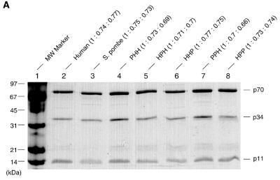
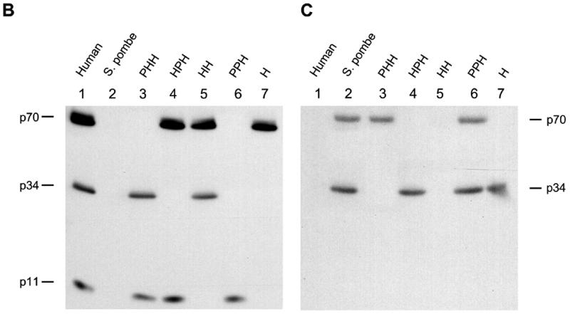
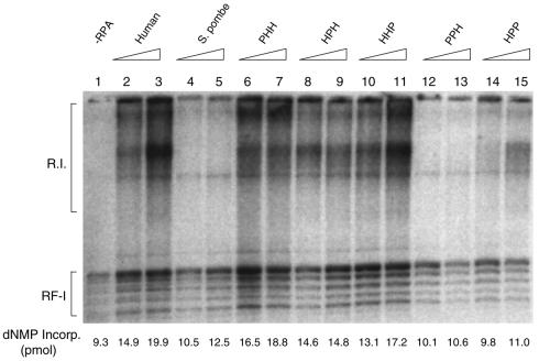
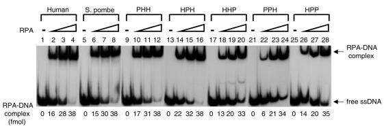
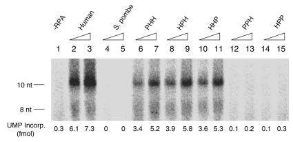
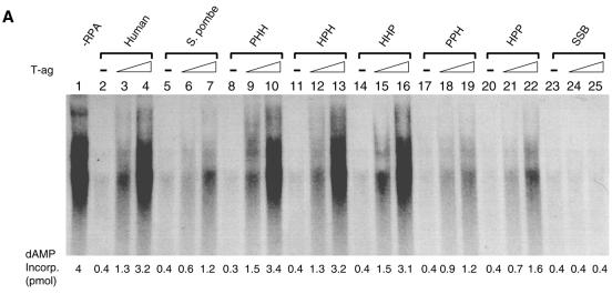
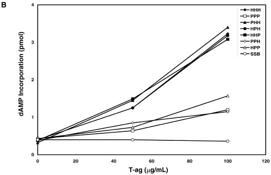
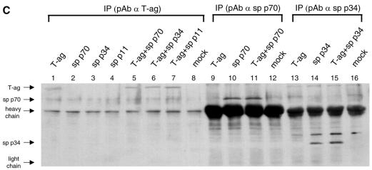
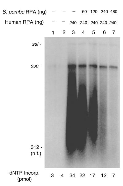
Replication protein A (RPA) is a three-subunit protein complex with multiple functions in DNA replication. Previous study indicated that human RPA (h-RPA) could not be replaced by Schizosaccharomyces pombe RPA (sp-RPA) in simian virus 40 (SV40) replication, suggesting that h-RPA may have a specific function in SV40 DNA replication. To understand the specificity of h-RPA in replication, we prepared heterologous RPAs containing the mixture of human and S.pombe subunits and compared these preparations for various enzymatic activities. Heterologous RPAs containing two human subunits supported SV40 DNA replication, whereas those containing only one human subunit poorly supported DNA replication, suggesting that RPA complex requires at least two human subunits to support its function in SV40 DNA replication. All heterologous RPAs effectively supported single-stranded (ss)DNA binding activity and an elongation of a primed DNA template catalyzed by DNA polymerase (pol) alpha and delta. A strong correlation between SV40 DNA replication activity and large tumor antigen (T-ag)-dependent RNA primer synthesis by pol alpha-primase complex was observed among the heterologous RPAs. Furthermore, T-ag showed a strong interaction with 70- and 34-kDa subunits from human, but poorly interacted with their S.pombe counterparts, indicating that the specificity of h-RPA is due to its role in RNA primer synthesis. In the SV40 replication reaction, the addition of increasing amounts of sp-RPA in the presence of fixed amount of h-RPA significantly reduced overall DNA synthesis, but increased the size of lagging strand, supporting a specific role for h-RPA in RNA primer synthesis. Together, these results suggest that the specificity of h-RPA in SV40 replication lies in T-ag-dependent RNA primer synthesis.


Figures

Figure 1
Figure 1
SDS–PAGE and western blot of heterologous RPA from h- and sp-RPA was purified using a previously described protocol (23) and analyzed by 12% SDS–PAGE (A) or immunoblotting procedure using mixtures of anti-p70, -p34, -p11 antibodies raised against h-RPA subunits (B) or the mixtures of anti-p70- and -p34 antibodies against sp-RPA subunits (C). The subunit stoichiometry of various RPA complex (p70: p34: p11) was measured by densitometric scanning and is indicated at the top of (A). The three-letter symbols at the top of the figure represent the source of RPA subunits [either from human (H) or from S.pombe (P)] in the order of p70, p34 and p11. For example, PHH represents RPA complex containing sp-p70, h-p34 and h-p11.
Figure 1
Figure 1
SDS–PAGE and western blot of heterologous RPA from h- and sp-RPA was purified using a previously described protocol (23) and analyzed by 12% SDS–PAGE (A) or immunoblotting procedure using mixtures of anti-p70, -p34, -p11 antibodies raised against h-RPA subunits (B) or the mixtures of anti-p70- and -p34 antibodies against sp-RPA subunits (C). The subunit stoichiometry of various RPA complex (p70: p34: p11) was measured by densitometric scanning and is indicated at the top of (A). The three-letter symbols at the top of the figure represent the source of RPA subunits [either from human (H) or from S.pombe (P)] in the order of p70, p34 and p11. For example, PHH represents RPA complex containing sp-p70, h-p34 and h-p11.
Figure 2
Figure 2
The effect of heterologous RPA on SV40 DNA replication reconstituted with purified proteins in vitro. Replication reactions contained SV40 pUC-ori+, 0.6 µg of SV40 T-ag and 100 µg of ammonium sulfate fraction (35–65%) from HeLa cell extracts. Indicated amounts of RPA (150 ng in lanes 2, 4, 6, 8, 10, 12 and 14, and 300 ng in lanes 3, 5, 7, 9, 11, 13 and 15) were added to the reaction. The composition of the RPA is indicated by the three-letter symbol at the top of the figure. Reaction mixtures were incubated for 90 min at 37°C, and the reaction products were analyzed by agarose gel electrophoresis (1.2%) and acid-insoluble radioactivity.
Figure 3
Figure 3
The ssDNA binding activity of various heterologous RPAs. Three concentrations (5, 10 and 20 ng) of RPA containing either the human or S.pombe subunit, as indicated at the top of the figure, were incubated with 100 fmol of 5′-32P-labeled (dT)50 for 15 min at room temperature. The RPA–DNA complexes were separated from free DNA by 5% PAGE (acrylamide:bisacrylamide = 79:1). For quantitation, the protein–DNA complex bands were excised and analyzed by liquid scintillation counting.
Figure 4
Figure 4
Effects of heterologous RPAs on RNA-primer synthesis during the initiation of SV40 DNA replication. Reaction mixtures containined 0.3 µg of SV40 pUC-ori+, 0.8 µg of SV40 T-ag, 1200 U of topoisomerase I, pol α (0.6 U)–primase (3 U) complex, and rNTPs but no dNTPs (see Materials and Methods for details). Where indicated, increasing amounts of RPA (150 ng in lanes 2, 4, 6, 8, 10, 12 and 14; 300 ng in lanes 3, 5, 7, 9, 11, 13 and 15) were added to the reaction. The composition of the RPA is indicated by the three-letter symbol at the top of the figure. After incubation at 37°C for 30 min, reaction products were processed as described in Materials and Methods and analyzed by 20% PAGE containing 7 M urea, 1 mM EDTA and 89 mM Tris–borate (pH 8.0).
Figure 5
Figure 5
Effect of heterologous RPA on the coupled DNA pol α–primase activity on ssM13 DNA. (A) The coupled DNA pol α–primase reactions containing 40 ng of unprimed M13 ssDNA template and 0.1 U of human pol α–primase complex were incubated at 37°C for 30 min in the presence of 0.4 µg RPA (except lane 1) and the increasing amount of T-ag (0 µg, lanes 1, 2, 5, 8, 11, 14, 17, 20 and 23; 0.4 µg, lanes 3, 6, 9, 12, 15, 18, 21 and 24; 0.8 µg, lanes 4, 7, 10, 13, 16, 19, 22 and 25). The heterologous RPAs used are indicated by the three-letter symbol. After incubation, reaction products were isolated as described in Materials and Methods and analyzed by 20% PAGE containing 7 M urea, 1 mM EDTA and 89 mM Tris–borate (pH 8.0). (B) For quantitation, regions containing reaction products were excised and analyzed by liquid scintillation counting.
Figure 5
Figure 5
Effect of heterologous RPA on the coupled DNA pol α–primase activity on ssM13 DNA. (A) The coupled DNA pol α–primase reactions containing 40 ng of unprimed M13 ssDNA template and 0.1 U of human pol α–primase complex were incubated at 37°C for 30 min in the presence of 0.4 µg RPA (except lane 1) and the increasing amount of T-ag (0 µg, lanes 1, 2, 5, 8, 11, 14, 17, 20 and 23; 0.4 µg, lanes 3, 6, 9, 12, 15, 18, 21 and 24; 0.8 µg, lanes 4, 7, 10, 13, 16, 19, 22 and 25). The heterologous RPAs used are indicated by the three-letter symbol. After incubation, reaction products were isolated as described in Materials and Methods and analyzed by 20% PAGE containing 7 M urea, 1 mM EDTA and 89 mM Tris–borate (pH 8.0). (B) For quantitation, regions containing reaction products were excised and analyzed by liquid scintillation counting.
Figure 6
Figure 6
Interaction of SV40 T-ag and RPA subunits in vivo. (A and B) Singly or coinfected with recombinant baculoviruses encoding T-ag and h-RPA subunit are indicated at top of panels. Lysates prepared from [35S]methionine-labeled insect cells were incubated with anti-T-ag antibody (A), anti-human p70 polyclonal antibody (B, lanes 1–4), or anti-human p34 polyclonal antibody (B, lanes 5–8). The immune complexes were precipitated with protein A–Sepharose and analyzed by 12% SDS–PAGE. (C) Singly or coinfected with recombinant baculoviruses encoding T-ag and sp-RPA subunit are indicated at top of panel. Lysates prepared from insect cells (without [35S]methionine) were incubated with anti-T-ag antibody (lanes 1–8), anti-sp-p70 polyclonal antibody (lanes 9–12), or anti-sp-p34 polyclonal antibody (lanes 13–16). The immune complexes were precipitated with protein A–Sepharose and analyzed by 12% SDS–PAGE followed by western blot using the mixture of anti-sp-p70 and sp-p34 polyclonal antibodies. Immunoglobulin heavy chain and light chain are indicated by arrows.
Figure 6
Figure 6
Interaction of SV40 T-ag and RPA subunits in vivo. (A and B) Singly or coinfected with recombinant baculoviruses encoding T-ag and h-RPA subunit are indicated at top of panels. Lysates prepared from [35S]methionine-labeled insect cells were incubated with anti-T-ag antibody (A), anti-human p70 polyclonal antibody (B, lanes 1–4), or anti-human p34 polyclonal antibody (B, lanes 5–8). The immune complexes were precipitated with protein A–Sepharose and analyzed by 12% SDS–PAGE. (C) Singly or coinfected with recombinant baculoviruses encoding T-ag and sp-RPA subunit are indicated at top of panel. Lysates prepared from insect cells (without [35S]methionine) were incubated with anti-T-ag antibody (lanes 1–8), anti-sp-p70 polyclonal antibody (lanes 9–12), or anti-sp-p34 polyclonal antibody (lanes 13–16). The immune complexes were precipitated with protein A–Sepharose and analyzed by 12% SDS–PAGE followed by western blot using the mixture of anti-sp-p70 and sp-p34 polyclonal antibodies. Immunoglobulin heavy chain and light chain are indicated by arrows.
Figure 7
Figure 7
Effect of sp-RPA on the size of replication products formed in the in vitro SV40 replication system. Reaction mixtures (40 µl) contained SV40 pUC-ori+, 0.6 µg of SV40 T-ag, 1200 U of topoisomerase I, pol α (0.1 U)–primase (0.5 U) complex, RF-C (0.1 µg), PCNA (0.2 µg) and pol δ (0.05 U), and the indicated amounts of sp- and h-RPA. After incubation at 37°C for 90 min, replication products were isolated and analyzed by 1.2% alkaline agarose gel electrophoresis. ssl and ssc indicate the position of single-stranded linear and single-stranded circular DNA, respectively.

Similar articles

Cited by

References

    1. Wobbe C.R., Weissbach,L., Borowiec,J.A., Dean,F.B., Murakami,Y., Bullock,P. and Hurwitz,J. (1987) Proc. Natl Acad. Sci. USA., 82, 5710–5714. - PubMed
    1. Fairman M.P. and Stillman,B. (1988) EMBO J., 7, 1211–1218. - PMC - PubMed
    1. Wold M.S. and Kelly,T.J. (1988) Proc. Natl Acad. Sci. USA, 85, 2523–2527. - PMC - PubMed
    1. Brill S.J. and Stillman,B. (1991) Genes Dev., 5, 1589–1600. - PubMed
    1. Erdile L.F., Heyer,W.-D., Kolodner,R. and Kelly,T.J. (1991) J. Biol. Chem., 266, 12090–12098. - PubMed

Publication types