. 2000 Dec 1;28(23):E103.
doi: 10.1093/nar/28.23.e103.

Mapping dispersed repetitive loci using semi-specific PCR cloning and somatic cell hybrid mapping

Affiliations

Mapping dispersed repetitive loci using semi-specific PCR cloning and somatic cell hybrid mapping

Y M Deng et al. Nucleic Acids Res. .

Abstract

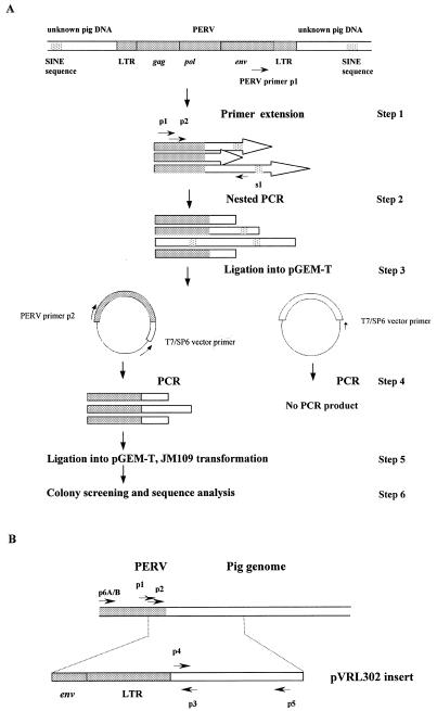
A simple and effective method based upon semi-specific PCR followed by cloning has been developed. Chromosomal mapping of the generated fragment on a somatic cell hybrid panel identifies the chromosomal position, and yields a unique sequence tag for the site. Using this method, the chromosomal location of one porcine endogenous retrovirus (PERV) was determined. The porcine genomic sequences were first amplified by PCR using a PERV-specific primer and a porcine short interspersed nuclear element (SINE)-specific primer. PCR products were cloned, and those sequences that contained PERV plus flanking regions were selected using a second round of PCR and cloning. Sequences flanking the PERV were determined and a PERV-B was physically mapped on porcine chromosome 17 using a somatic hybrid panel. The general utility of the method was subsequently demonstrated by locating PERVs in the genome of PERV infected human 293 cells. This method obviates the need for individual library construction or linker/adaptor ligation, and can be used to quickly locate individual sites of moderately repeated, dispersed DNA sequences in any genome.


Figures

Figure 1
Figure 1
Amplification and cloning of regions flanking PERVs in the pig genome. The shaded bars represent a PERV sequence, the open bars represent the unknown pig genome sequences flanking the PERV, and the cross-hatched area represents SINE sequences. The long arrows represent primer extension products, and short arrows the PCR primers. (A) Overall procedure. Step 1: primer extension on pig DNA using PERV-specific primer (p1). Step 2: PCR using PERV and SINE primers (p1, p2 and s1). Step 3: PCR products are cloned into a suitable vector (pGEM-T). Step 4: PCR of the ligation mixture using PERV (p2) and vector primers (T7/SP6). Step 5: PCR products are cloned into a suitable vector (pGEM-T). Step 6: screening of colonies for PERV positive clones and sequence confirmation. (B) Locations of PCR primers on the region containing part of PERV with its downstream flanking sequence. The fragment that was cloned in pVRL302 is shown underneath. (C) Amplification products from Large White × Landrace pig DNA. PCR was performed using primers p1 and p3 (lane 2), PERV-A specific primer p6A and primer p3 (lane 3), PERV-B specific primer p6B and primer p3 (lane 5). Water controls were included for each PCR (lanes 1, 4 and 6). M, molecular weight marker (pGEM DNA marker).
Figure 1
Figure 1
Amplification and cloning of regions flanking PERVs in the pig genome. The shaded bars represent a PERV sequence, the open bars represent the unknown pig genome sequences flanking the PERV, and the cross-hatched area represents SINE sequences. The long arrows represent primer extension products, and short arrows the PCR primers. (A) Overall procedure. Step 1: primer extension on pig DNA using PERV-specific primer (p1). Step 2: PCR using PERV and SINE primers (p1, p2 and s1). Step 3: PCR products are cloned into a suitable vector (pGEM-T). Step 4: PCR of the ligation mixture using PERV (p2) and vector primers (T7/SP6). Step 5: PCR products are cloned into a suitable vector (pGEM-T). Step 6: screening of colonies for PERV positive clones and sequence confirmation. (B) Locations of PCR primers on the region containing part of PERV with its downstream flanking sequence. The fragment that was cloned in pVRL302 is shown underneath. (C) Amplification products from Large White × Landrace pig DNA. PCR was performed using primers p1 and p3 (lane 2), PERV-A specific primer p6A and primer p3 (lane 3), PERV-B specific primer p6B and primer p3 (lane 5). Water controls were included for each PCR (lanes 1, 4 and 6). M, molecular weight marker (pGEM DNA marker).
Figure 2
Figure 2
Schematic illustration of the location of the PCR product on the chromosome. PCR product with expected size was generated from two out of the 27 porcine–rodent somatic hybrid clones (indicated by +). The proportion of pig chromosome 17 retained in each of the 27 somatic hybrid clones is shown as a solid bar.

References

    1. Frankel W.N., Stoye,J.P., Taylor,B.A. and Coffin,J.M. (1990) Genetics, 124, 221–236. - PMC - PubMed
    1. Tereba A. (1983) Curr. Topics Microbiol. Immunol., 107, 29–50. - PubMed
    1. Rogel-Gaillard C., Bourgeaux,N., Billault,A., Vaiman,M. and Chardon,P. (1999) Cytogenet. Cell Genet., 85, 205–211. - PubMed
    1. Rosenthal A. and Jones,D.S.C. (1990) Nucleic Acids Res., 18, 3095–3096. - PMC - PubMed
    1. Siebert P.D., Chenchik,A., Kellogg,D.E., Lukyanov,K.A. and Lukyanov,S.A. (1995) Nucleic Acids Res., 23, 1087–1088. - PMC - PubMed

Publication types

Associated data