

Crystal structures of photosynthetic reaction center and high-potential iron-sulfur protein from Thermochromatium tepidum: thermostability and electron transfer
- PMID: 11095707
- PMCID: PMC17615
- DOI: 10.1073/pnas.240224997
Crystal structures of photosynthetic reaction center and high-potential iron-sulfur protein from Thermochromatium tepidum: thermostability and electron transfer
Abstract
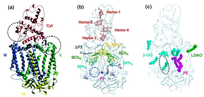
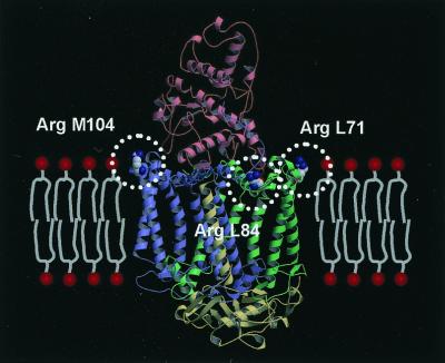
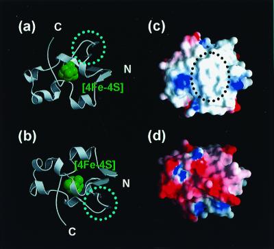
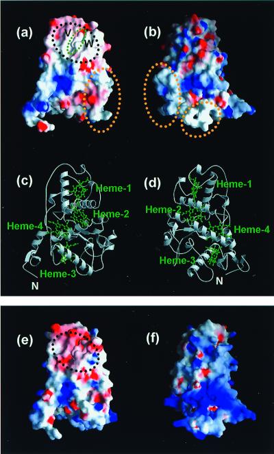
The reaction center (RC) of photosynthetic bacteria is a membrane protein complex that promotes a light-induced charge separation during the primary process of photosynthesis. In the photosynthetic electron transfer chain, the soluble electron carrier proteins transport electrons to the RC and reduce the photo-oxidized special-pair of bacteriochlorophyll. The high-potential iron-sulfur protein (HiPIP) is known to serve as an electron donor to the RC in some species, where the c-type cytochrome subunit, the peripheral subunit of the RC, directly accepts electrons from the HiPIP. Here we report the crystal structures of the RC and the HiPIP from Thermochromatium (Tch.) tepidum, at 2.2-A and 1.5-A resolution, respectively. Tch. tepidum can grow at the highest temperature of all known purple bacteria, and the Tch. tepidum RC shows some degree of stability to high temperature. Comparison with the RCs of mesophiles, such as Blastochloris viridis, has shown that the Tch. tepidum RC possesses more Arg residues at the membrane surface, which might contribute to the stability of this membrane protein. The RC and the HiPIP both possess hydrophobic patches on their respective surfaces, and the HiPIP is expected to interact with the cytochrome subunit by hydrophobic interactions near the heme-1, the most distal heme to the special-pair.
Figures
Similar articles
-
Crystal structure of a photosynthetic LH1-RC in complex with its electron donor HiPIP.Nat Commun. 2021 Feb 17;12(1):1104. doi: 10.1038/s41467-021-21397-9. Nat Commun. 2021. PMID: 33597527 Free PMC article.
-
Structural and functional studies on the tetraheme cytochrome subunit and its electron donor proteins: the possible docking mechanisms during the electron transfer reaction.Photosynth Res. 2005;85(1):87-99. doi: 10.1007/s11120-004-2416-5. Photosynth Res. 2005. PMID: 15977061 Review.
-
Structure of the H subunit of the photosynthetic reaction center from the thermophilic purple sulfur bacterium, Thermochromatium tepidum Implications for the specific binding of the lipid molecule to the membrane protein complex.Eur J Biochem. 2001 May;268(9):2652-7. doi: 10.1046/j.1432-1327.2001.02158.x. Eur J Biochem. 2001. PMID: 11322886
-
Interaction site for high-potential iron-sulfur protein on the tetraheme cytochrome subunit bound to the photosynthetic reaction center of Rubrivivax gelatinosus.Biochemistry. 1999 Mar 9;38(10):2861-5. doi: 10.1021/bi982747w. Biochemistry. 1999. PMID: 10074337
-
Structural basis of bacterial photosynthetic reaction centers.J Biochem. 2001 Sep;130(3):319-29. doi: 10.1093/oxfordjournals.jbchem.a002989. J Biochem. 2001. PMID: 11530006 Review.
Cited by
-
Multiscale Simulations of Biological Membranes: The Challenge To Understand Biological Phenomena in a Living Substance.Chem Rev. 2019 May 8;119(9):5607-5774. doi: 10.1021/acs.chemrev.8b00538. Epub 2019 Mar 12. Chem Rev. 2019. PMID: 30859819 Free PMC article.
-
Crystal structure of a photosynthetic LH1-RC in complex with its electron donor HiPIP.Nat Commun. 2021 Feb 17;12(1):1104. doi: 10.1038/s41467-021-21397-9. Nat Commun. 2021. PMID: 33597527 Free PMC article.
-
Which side of the pi-macrocycle plane of (bacterio)chlorophylls is favored for binding of the fifth ligand?Photosynth Res. 2002;74(1):1-10. doi: 10.1023/A:1020816128794. Photosynth Res. 2002. PMID: 16228541
-
Structural and functional studies on the tetraheme cytochrome subunit and its electron donor proteins: the possible docking mechanisms during the electron transfer reaction.Photosynth Res. 2005;85(1):87-99. doi: 10.1007/s11120-004-2416-5. Photosynth Res. 2005. PMID: 15977061 Review.
-
The structure and function of the cytochrome c2: reaction center electron transfer complex from Rhodobacter sphaeroides.Photosynth Res. 2005;85(1):101-14. doi: 10.1007/s11120-005-1368-8. Photosynth Res. 2005. PMID: 15977062 Review.
References
-
- Madigan M T. Science. 1984;225:313–315. - PubMed
-
- Madigan M T. Int J Syst Bacteriol. 1986;36:222–227.
-
- Nozawa T, Madigan M T. J Biochem (Tokyo) 1991;110:588–594. - PubMed
-
- Deisenhofer J, Epp O, Miki K, Huber R, Michel H. J Mol Biol. 1984;180:385–398. - PubMed
-
- Deisenhofer J, Epp O, Miki K, Huber R, Michel H. Nature (London) 1985;318:618–624. - PubMed
Publication types
MeSH terms
Substances
Associated data
- Actions
- Actions
LinkOut - more resources
Full Text Sources
Molecular Biology Databases
