

doi: 10.1073/pnas.240305897.
Caspase-1 and -3 are sequentially activated in motor neuron death in Cu,Zn superoxide dismutase-mediated familial amyotrophic lateral sclerosis
Affiliations
- PMID: 11095709
- PMCID: PMC17673
- DOI: 10.1073/pnas.240305897
Item in Clipboard
Caspase-1 and -3 are sequentially activated in motor neuron death in Cu,Zn superoxide dismutase-mediated familial amyotrophic lateral sclerosis
Proc Natl Acad Sci U S A.
.
Abstract
Familial amyotrophic lateral sclerosis-linked mutations in copper-zinc superoxide dismutase cause motor neuron death through one or more acquired toxic properties. An early event in the mechanism of toxicity from such mutants is now demonstrated to be activation of caspase-1. Neuronal death, however, follows only after months of chronic caspase-1 activation concomitantly with activation of the executioner caspase-3 as the final step in the toxic cascade. Thus, a common toxicity of mutant SOD1 is a sequential activation of at least two caspases, caspase-1 that acts slowly as a chronic initiator and caspase-3 acting as the final effector of cell death.
Figures
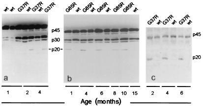
Caspase-1 cleavage appears early in the lifespan of G37R- and
G85R-transgenic mice. Immunoblots of spinal cord extracts from G37R
(a) and G85R (b) mice probed with a
polyclonal antibody against both the inactive (p45) pro-enzyme and the
active (p20) caspase-1. In mice over-expressing the wt human SOD1
(hSOD1) protein, the antibody only recognized the p45 inactive and the
p30 intermediate form. (c) Caspase-1 cleavage in G37R
mice detected with the polyclonal antibody used for the cell culture
experiments. wt indicates mice overexpressing wt hSOD1.
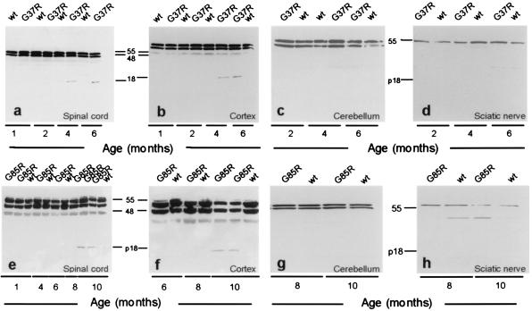
Caspase-3 is activated late in mutant SOD1-transgenic mouse spinal cord
and cortex but not in cerebellum or sciatic nerve. Immunoblots of
spinal cord and cortex extracts from G37R (a and
b) and G85R (e and
f) mice probed with the CM1 antibody. No
immunostaining is detected early in life. Staining for the p18 fragment
appears at 4 months in the G37R mice and at 10 months in the G85R mice
but not in littermate nontransgenic controls (wt). Activation of
caspase-3 is tissue-specific; no active fragment appears in cerebellum
or sciatic nerve of either G37R (c and d)
or G85R (g and h) mice. Each gel was
loaded with 30 μg/lane.
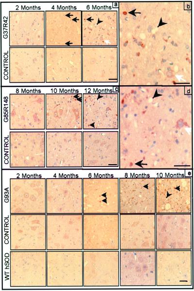
Caspase-3 immunoreactivity appears ≈2 months before onset and
increases with age in the spinal cord anterior horn of all mutant SOD1
mouse lines tested. Immunostaining for active caspase-3 (brown
precipitate) in G37R (a and b) and G85R
(c and d) mice and their nontransgenic
littermate controls. (b and d) Increased
magnification of a and c (end stage).
(e) Active caspase-3 immunoreactivity in G93A mouse,
nontransgenic littermate control, and wt hSOD1-expressing mouse spinal
cord. Black arrows indicate active caspase-3-positive inclusions. White
arrows indicate caspase-3-positive motor neurons. Arrowheads indicate
vacuoles. (Bars = 25 μm.)
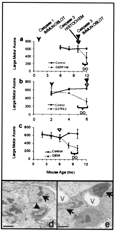
Caspase-1 and caspase-3 activation precedes loss of large motor
axons and the appearance of apoptotic morphology in all mutant
SOD1 mouse lines tested. Axon counts from the L5 motor root of the
spinal cord for G85R (a), G37R (b), and
G93A (c) mice (dotted lines) and their littermate
nontransgenic controls (solid lines) at various ages. The appearance of
activated caspase-1 in immunoblot is indicated by the leftmost
arrowhead in a and b. The appearance of
activated caspase-3 in immunoblot is indicated by solid triangles. The
appearance of activated caspase-3 in immunohistochemistry is indicated
by open triangles in a, b, and
c. Axon counts are averages from three to five animals
of each genotype and age. Horizontal brackets indicate the age of
disease onset (DO). Error bars are the SD of the data.
(d and e) Electron microscopy images show
apoptotic changes within the ventral horn of 8-mo-old
G93A-mutant mice. Arrows indicate condensed chromatin within nuclei.
Vacuoles are marked with a V. Filaments within neurons are indicated
with an F. (Bars in d and e = 2
μm.)
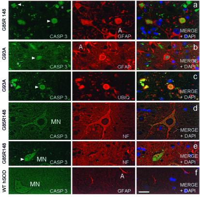
Activated caspase-3 appears within the neuronal and glial
inclusions present in mutant SOD1 mouse spinal cords. Double
immunofluorescence of spinal cord anterior horn using CM1 antibody
(Casp 3 in the Left panels) and either GFAP antibody to
stain astrocytes, ubiquitin antibody (UBIQ) to stain inclusions, or an
antibody recognizing nonphosphorylated neurofilaments (NF) to visualize
neurons. Right panels depict the merged image from the
first two channels with DAPI to show the position of nuclei. Activated
caspase-3-positive inclusions (arrowheads) within astrocytes (A)
and/or extracellular debris (*) are common features within
mutant G85R (a) and G93A-mutant (b)
spinal cords. (c) Activated caspase-3 colocalizes with
ubiquitin-positive inclusions in G93A-mutant mice. Activated caspase-3
immunoreactivity is present within motor neurons (MN)
(d) and motor neuron inclusions (e)
(arrow) of mutant G85R mice. (f) Activated
caspase-3 is absent from 10-mo-old wt hSOD1-transgenic mice whereas
astrocytic GFAP staining is normal. (Bar = 12.5 μm.)
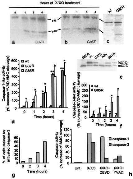
Caspase-1 is activated early in differentiated mouse
neuroblastoma N2a cells. Immunoblots showing caspase-1 activation in
differentiated G37R (a) or G85R (b)
positive N2a exposed to X/XO for 0, 1, 3, or 4 h.
(c) Immunoblot of lysates from wt hSOD1-expressing cells
(left) or G85R-positive cells (right) 4 h after X/XO treatment.
(d) YVAD-AMC cleavage was measured as described and is
reported relative to the cleavage induced by lysates of untreated (time
0) wt hSOD1-positive cultures. Data are the mean ± SD of three
independent experiments for the G85R-positive cells and of two
independent experiments assayed in duplicate for the G37R and wt hSOD1
expressing cells. Asterisks (*, P < 0.05;
**, P < 0.01) indicate significant
differences between groups. (e) Immunoblot showing
levels of wt and mutant SODs in N2a cell lines by using an antibody
that recognizes human and mouse SOD1 equally. The hSOD1 protein
migrates more slowly than the mouse, only the G85R mutant comigrates
with the mouse SOD1. (f) Caspase-3 activity
recorded as DEVD-AMC cleavage by cell lysates relative to the cleavage
induced by lysates of untreated wt hSOD1-positive cells. Data are the
mean ± SD of three independent experiments. Asterisks (*,
P < 0.05) indicate a statistical difference
between groups. (g) Percentage of cells that stained
positive for activated caspase-3 at different times after X/XO
treatment. (h) YVAD-AMC cleavage and DEVD-AMC cleavage
measured in lysates from G85R cells treated with X/XO in the presence
of caspase-1-and-3 inhibitors. Data are the mean ± SD of six
replicates. Fluorescence emitted from the fluorogenic substrate is
expressed relative to that emitted from untreated cells.
References
-
- Rosen D R, Siddique T, Patterson D, Figlewicz D A, Sapp P, Hentati A, Donaldson D, Goto J, O'Regan J P, Deng H X, et al. Nature (London) 1993;362:59–62. - PubMed
-
- Cleveland D W. Neuron. 1999;24:515–20. - PubMed
-
- Reaume A, Elliott J, Hoffman E, Kowall N, Ferrante R, Siwek D, Wilcox H, Flood D, Beal M, Brown, et al. Nat Genet. 1996;13:43–47. - PubMed
-
- Gurney M E, Pu H, Chiu A Y, Dal Canto M C, Polchow C Y, Alexander D D, Caliendo J, Hentati A, Kwon Y W, Deng H-X, et al. Science. 1994;264:1772–1775. - PubMed
-
- Wong P, Pardo C, Borchelt D, Lee M, Copeland N, Jenkins N, Sisodia S, Cleveland D, Price D. Neuron. 1995;14:1105–1116. - PubMed
Publication types
MeSH terms
Substances
Grants and funding
LinkOut - more resources
Full Text Sources
Other Literature Sources
Medical
Research Materials
Miscellaneous
