Adeno-associated virus vector carrying human minidystrophin genes effectively ameliorates muscular dystrophy in mdx mouse model
- PMID: 11095710
- PMCID: PMC17641
- DOI: 10.1073/pnas.240335297
Adeno-associated virus vector carrying human minidystrophin genes effectively ameliorates muscular dystrophy in mdx mouse model
Abstract
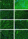
Duchenne muscular dystrophy (DMD) is the most common and lethal genetic muscle disorder, caused by recessive mutations in the dystrophin gene. One of every 3,500 males suffers from DMD, yet no treatment is currently available. Genetic therapeutic approaches, using primarily myoblast transplantation and adenovirus-mediated gene transfer, have met with limited success. Adeno-associated virus (AAV) vectors, although proven superior for muscle gene transfer, are too small (5 kb) to package the 14-kb dystrophin cDNA. Here we have created a series of minidystrophin genes (<4.2 kb) under the control of a muscle-specific promoter that readily package into AAV vectors. When injected into the muscle of mdx mice (a DMD model), two of the minigenes resulted in efficient and stable expression in a majority of the myofibers, restoring the missing dystrophin and dystrophin-associated protein complexes onto the plasma membrane. More importantly, this AAV treatment ameliorated dystrophic pathology in mdx muscle and led to normal myofiber morphology, histology, and cell membrane integrity. Thus, we have defined minimal functional dystrophin units and demonstrated the effectiveness of using AAV to deliver the minigenes in vivo, offering a promising avenue for DMD gene therapy.
Figures


Comment in
-
The dystrophin-associated glycoprotein complex: what parts can you do without?Proc Natl Acad Sci U S A. 2000 Dec 5;97(25):13464-6. doi: 10.1073/pnas.011510597. Proc Natl Acad Sci U S A. 2000. PMID: 11095702 Free PMC article. No abstract available.
Similar articles
-
[Adeno-associated virus vector carrying human minidystrophin gene SMCKA3999 effectively ameliorates dystrophic pathology in mdx model mice].Zhonghua Yi Xue Za Zhi. 2003 Sep 10;83(17):1513-6. Zhonghua Yi Xue Za Zhi. 2003. PMID: 14521733 Chinese.
-
Adeno-associated virus vector-mediated minidystrophin gene therapy improves dystrophic muscle contractile function in mdx mice.Hum Gene Ther. 2002 Aug 10;13(12):1451-60. doi: 10.1089/10430340260185085. Hum Gene Ther. 2002. PMID: 12215266
-
A canine minidystrophin is functional and therapeutic in mdx mice.Gene Ther. 2008 Aug;15(15):1099-106. doi: 10.1038/gt.2008.70. Epub 2008 Apr 24. Gene Ther. 2008. PMID: 18432277
-
[Gene therapy for muscular dystrophy].No To Hattatsu. 2004 Mar;36(2):117-23. No To Hattatsu. 2004. PMID: 15031985 Review. Japanese.
-
Recombinant adeno-associated viral (rAAV) vectors as therapeutic tools for Duchenne muscular dystrophy (DMD).Gene Ther. 2004 Oct;11 Suppl 1:S109-21. doi: 10.1038/sj.gt.3302379. Gene Ther. 2004. PMID: 15454965 Review.
Cited by
-
AAV-mediated overexpression of human α7 integrin leads to histological and functional improvement in dystrophic mice.Mol Ther. 2013 Mar;21(3):520-5. doi: 10.1038/mt.2012.281. Epub 2013 Jan 15. Mol Ther. 2013. PMID: 23319059 Free PMC article.
-
Multi-parametric MRI at 14T for muscular dystrophy mice treated with AAV vector-mediated gene therapy.PLoS One. 2015 Apr 9;10(4):e0124914. doi: 10.1371/journal.pone.0124914. eCollection 2015. PLoS One. 2015. PMID: 25856443 Free PMC article.
-
Enhancement of muscle gene delivery with pseudotyped adeno-associated virus type 5 correlates with myoblast differentiation.J Virol. 2001 Aug;75(16):7662-71. doi: 10.1128/JVI.75.16.7662-7671.2001. J Virol. 2001. PMID: 11462038 Free PMC article.
-
Problems and solutions in myoblast transfer therapy.J Cell Mol Med. 2001 Jan-Mar;5(1):33-47. doi: 10.1111/j.1582-4934.2001.tb00136.x. J Cell Mol Med. 2001. PMID: 12067449 Free PMC article. Review.
-
Viral-mediated gene therapy for the muscular dystrophies: successes, limitations and recent advances.Biochim Biophys Acta. 2007 Feb;1772(2):243-62. doi: 10.1016/j.bbadis.2006.09.007. Epub 2006 Sep 26. Biochim Biophys Acta. 2007. PMID: 17064882 Free PMC article. Review.
References
-
- Kunkel L M. Nature (London) 1986;322:73–77. - PubMed
-
- Koenig M, Hoffman E P, Bertelson C J, Monaco A P, Feener C, Kunkel L M. Cell. 1987;50:509–517. - PubMed
-
- Hoffman E P, Brown R H, Jr, Kunkel L M. Cell. 1987;51:919–928. - PubMed
-
- Koenig M, Kunkel L M. J Biol Chem. 1990;265:4560–4566. - PubMed
-
- Gussoni E, Blau H M, Kunkel L M. Nat Med. 1997;3:970–977. - PubMed
Publication types
MeSH terms
Substances
Grants and funding
LinkOut - more resources
Full Text Sources
Other Literature Sources
Medical
Miscellaneous

