. 2000 Dec 5;97(25):13619-24.
doi: 10.1073/pnas.240345197.

Glucose depletion causes haploid invasive growth in yeast

Affiliations

Glucose depletion causes haploid invasive growth in yeast

P J Cullen et al. Proc Natl Acad Sci U S A. .

Abstract

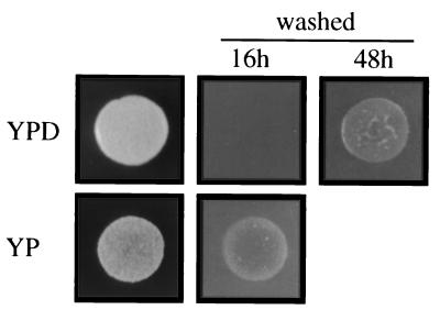
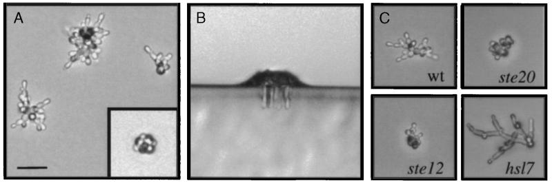
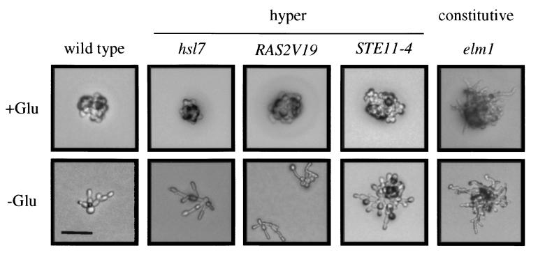
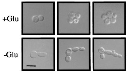
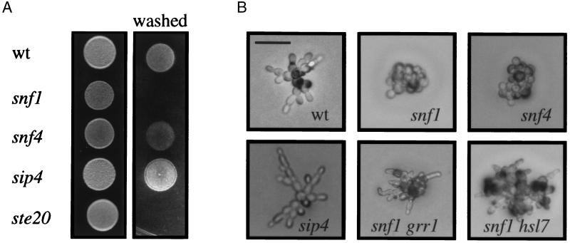
Haploid yeast invades solid agar in response to nutrient limitation. To decipher the cues that underlie invasion, we have developed a single cell invasive growth assay. Using this assay, as well as the traditional plate-washing assay, we show that invasive growth occurs in response to glucose depletion. In the absence of glucose (or other fermentable sugar), individual cells adopted a nonaxial budding pattern and elongated morphology within the first cell divisions, and invasion into the agar was observed in microcolonies containing as few as 10 cells. In support of this observation, we found that glucose suppressed the hyperinvasive growth morphology of STE11-4, pbs2, hsl7, and RAS2V19 mutations. In addition, removal of glucose from YPD medium caused constitutive invasion in wild-type cells. We tested glucose control proteins for a role in invasion and found that Snf1, a protein required for derepression of glucose-repressed genes, was required for invasive growth. The transcription factor Sip4, which interacts with Snf1 and is induced during the diauxic shift, had an inhibitory role on invasive growth, suggesting that multiple mechanisms are required for glucose depletion-dependent invasion.


Figures

Figure 1
Figure 1
Removal of glucose causes constitutive invasion. Equal concentrations of cells were spotted onto YPD (plus glucose) or YP medium (minus glucose), incubated for 16 h and 48 h, and photographed. Growth of cells on YP presumably occurred using poor carbon sources present in the yeast extract or agar.
Figure 2
Figure 2
Single cell invasive growth assay. (A) Cells of the Σ1278b background were spread onto SC medium and incubated for 24 h. Microcolonies were visualized by light microscopy at ×200. (Inset) Typical example of a microcolony formed in the presence of glucose at the same magnification. (B) Agar invasion on medium lacking glucose. Wild-type cells were spread onto SC medium and incubated for 24 h at 25°C. Plates were cut with a razor blade, and sections were set perpendicular to the plane of invasion for direct visualization by light microscopy. (C) Invasive growth mutants have phenotypes on medium lacking glucose. Equal concentrations of cells were spread onto medium lacking glucose. Mutant genotypes are as follows: Upper Left, wild type; Upper Right, ste20; Lower Left, ste12; and Lower Right, hsl7. A, B, and C are at the same scale, with the scale bar in A representing 40 μm.
Figure 3
Figure 3
Suppression of hyperinvasive growth mutant phenotypes by glucose. Equal concentrations of cells were grown for 16 h on SCD or SC medium. A 5-μl spot of 1 M glucose was placed onto a filter at the center of SC medium to allow observation of both yf and ff cell types. The RAS2V19 allele was compared on YP and YPD medium because the invasive growth phenotype was more apparent on rich medium. All pictures were at the same scale, with the bar in the lower left-hand corner representing 40 μm.
Figure 4
Figure 4
Cells grown with or without glucose have strikingly different morphologies in the first cell divisions. Cells were grown on SCD or SC medium, scraped off the plates, centrifuged, and photographed. Typical examples of microcolonies were shown. All pictures were at the same scale, with the bar in the Lower Left corner representing 10 μm.
Figure 5
Figure 5
Snf1 and Sip4 are required for proper invasion. (A) Equal concentrations of cells were spotted onto YPD medium and grown for 3 days at 30°C. The plates were photographed, washed, and photographed again. (B) Equal concentrations of cells were spread onto SC medium, incubated for 16 h at 25°C, and photographed. For B, all pictures were taken at the same scale, with the bar in the Upper Left corner representing 40 μm.

Comment in

Similar articles

Cited by

References

    1. Silar P, Daboussi M J. Trends Genet. 1999;15:141–145. - PubMed
    1. Gimeno C J, Ljungdahl P O, Styles C A, Fink G R. Cell. 1992;68:1077–1090. - PubMed
    1. Madhani H D, Fink G R. Trends Cell Biol. 1998;8:348–353. - PubMed
    1. Banuett F. Microbiol Mol Biol Rev. 1998;62:249–274. - PMC - PubMed
    1. Levin D E, Errede B. Curr Opin Cell Biol. 1995;7:197–202. - PubMed

Publication types

MeSH terms