. 2000 Dec 5;97(25):13537-42.
doi: 10.1073/pnas.240460997.

A continuous assay for DNA cleavage: the application of "break lights" to enediynes, iron-dependent agents, and nucleases

Affiliations

A continuous assay for DNA cleavage: the application of "break lights" to enediynes, iron-dependent agents, and nucleases

J B Biggins et al. Proc Natl Acad Sci U S A. .

Abstract

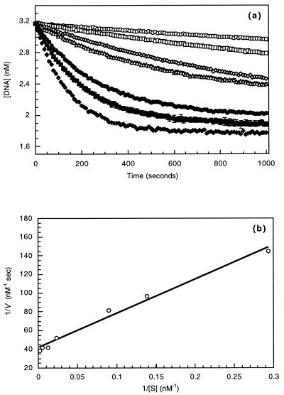
Although extensive effort has been applied toward understanding the mechanism by which enediynes cleave DNA, a continuous assay for this phenomenon is still lacking. In fact, with the exception of assays for DNase, continuous assays for most DNA cleavage events are unavailable. This article describes the application of "molecular break lights" (a single-stranded oligonucleotide that adopts a stem-and-loop structure and carries a 5'-fluorescent moiety, a 3'-nonfluorescent quenching moiety, and an appropriate cleavage site within the stem) to develop the first continuous assay for cleavage of DNA by enediynes. Furthermore, the generality of this approach is demonstrated by using the described assay to directly compare the DNA cleavage by naturally occurring enediynes [calicheamicin and esperamicin), non-enediyne small molecule agents (bleomycin, methidiumpropyl-EDTA-Fe(II), and EDTA-Fe(II]), as well as the restriction endonuclease BamHI. Given the simplicity, speed, and sensitivity of this approach, the described methodology could easily be extended to a high throughput format and become a new method of choice in modern drug discovery to screen for novel protein-based or small molecule-derived DNA cleavage agents.


Figures

Figure 1
Figure 1
Nonenzymatic DNA-cleaving agents: calicheamicin γ1I from M. echinospora (1), esperamicin A1 from A. verrucosospora (2), bleomycin from S. verticillus (3), MPE–Fe(II) (4), and EDTA–Fe(II) (5).
Figure 2
Figure 2
A schematic diagram of molecular beacons, molecular break lights, and the specific break lights used in this study. The solid lines represent covalent bonds, dashed lines represent hydrogen bonding, letters represent arbitrary bases, the gray shaded ball represents the fluorophore (FAM), the black ball represents the corresponding quencher (DABCYL), and the dashed wedges represent fluorescence. (a) Principle of operation of molecular beacons. Target hybridization leads to a separation of the fluorophore–quencher pair and a corresponding fluorescent signal. (b) Principle of operation of molecular break lights. Cleavage of the stem by an enzymatic or nonenzymatic nuclease activity results in the separation of the fluorophore–quencher pair and a corresponding fluorescent signal. (c) Molecular break lights used in this study. The stem of break light A contains a preferred calicheamicin recognition site (in bold), and the stem of break light B carries the BamHI recognition site (in bold). The predicted cleavage sites are illustrated by arrows.
Figure 3
Figure 3
The demonstration of molecular break light specificity and general proof of principle. The observed change in fluorescence intensity over time of an assay containing 3.2 nM break light at 37°C. (a) Break light A with 100 units of BamHI (□), break light B with 100 units of BamHI (○), and break light B without enzyme (●) (10 mM Tris⋅HCl/50 mM NaCl/10 mM MgCl2/1 mM DTT, pH 7.9; λEx = 485 nm, λEm = 517 nM). (b) Break light A with and 10 units of DNaseI (□), break light B with 10 units of DNaseI (○), and break light A without enzyme (●) (40 mM Tris⋅HCl/10 mM MgSO4/1 mM CaCl2, pH 8.0; λEx = 485 nm, λEm = 517 nM).
Figure 4
Figure 4
The determination of BamHI steady-state kinetic parameters using break light B. (a) The observed change in fluorescence intensity over time of an assay containing a constant 3.2 nM break light B at 37°C (6 mM Tris⋅HCl/100 mM NaCl/6 mM MgCl2/1 mM DTT, pH 7.5; λEx = 485 nm, λEm = 517 nM), BamHI (10 units), and varying nonlabeled substrate oligonucleotide. Total substrate concentrations (including break light): 389 nM (○), 196 nM (□), 81 nM (⋄), 42 nM (▵), 11 nM (●), 7.5 nM (■), and 3.4 nM (⧫). (b) Lineweaver–Burke plot from a after correction for the carrier dilution effect.
Figure 5
Figure 5
Cleavage of break light A by calicheamicin and esperamicin. The observed DNA cleavage over time of an assay containing 3.2 nM break light A at 37°C (40 mM Tris⋅HCl, pH 7.5; λEx = 485 nm, λEm = 517 nM), DTT (50 μM) and varied enediyne. (a) Calicheamicin concentrations: 31.7 nM (○), 15.9 nM (□), 3.2 nM (⋄), 1.6 nM (▵), 0.78 nM (●), and 0.31 nM (■). (b) Esperamicin concentrations: 31.7 nM (○), 15.9 nM (□), 3.2 nM (⋄), 1.6 nM (▵), 0.78 nM (●), 0.31 nM (■), and 0.15 nM (♦).
Figure 6
Figure 6
Cleavage of break light A by Fe(II)-dependent agents. (A) The observed DNA cleavage over time of an assay containing a constant 3.2 nM break light A at 37°C (50 mM sodium phosphate/2.5 mM ascorbate, pH 7.5; λEx = 485 nm, λEm = 517 nM) and varied bleomycin. Bleomycin concentrations: 200 nM (○), 100 nM (□), 50 nM (⋄), 25 nM (▵), 12.5 nM (●), 5 nM (■), and 2.5 nM (▴). (B) The observed DNA cleavage over time of an assay containing a constant 3.2 nM break light A at 37°C (40 mM Tris⋅HCl/2.5 mM ascorbate, pH 7.5; λEx = 485 nm, λEm = 517 nM) and varied MPE. Fe(II) concentrations: 8 μM (○), 4 μM (□), 2 μM (⋄), 1 μM (▵), 500 nM (●), 250 nM (■), and 125 nM (▴). (C) The observed DNA cleavage over time of an assay containing a constant 32 nM break light A at 37°C (40 mM Tris⋅HCl/2.5 mM ascorbate, pH 7.5; λEx = 485 nm, λEm = 517 nM) and varied MPE. Fe(II) concentrations: 50 nM (○), 125 nM (□), 250 nM (⋄), 500 nM (▵), 1 μM (●), and 2 μM (■). (D) The observed DNA cleavage over time of an assay containing a constant 32 nM break light A at 37°C (40 mM Tris⋅HCl/2.5 mM ascorbate, pH 7.5; λEx = 485 nm, λEm = 517 nM) and varied Fe(II)–EDTA. Fe(II) concentrations: 12.5 μM (○), 6.3 μM (□), 3.1 μM (⋄), and 1.3 μM (▵).

Similar articles

Cited by

References

    1. Thorson, J. S., Sievers, E. L., Ahlert, J., Shepard, E., Onwueme, K. C. & Ruppen, M. (2000) Curr. Pharm. Des., in press. - PubMed
    1. Thorson J S, Shen B, Whitwam R E, Liu W, Li Y, Ahlert J. Bioorgan Chem. 1999;27:172–188.
    1. Borders D B, Doyle T W. Enediyne Antibiotics as Antitumor Agents. New York: Dekker; 1995.
    1. Smith A L, Nicolaou K C. J Med Chem. 1996;39:2103–2117. - PubMed
    1. Nicolaou K C, Smith A L, Yue E W. Proc Natl Acad Sci USA. 1993;90:5881–5888. - PMC - PubMed

Publication types