. 2000 Dec 5;97(25):13602-7.
doi: 10.1073/pnas.250471997.

Structural insights into the binding of cardiac glycosides to the digitalis receptor revealed by solid-state NMR

Affiliations

Structural insights into the binding of cardiac glycosides to the digitalis receptor revealed by solid-state NMR

D A Middleton et al. Proc Natl Acad Sci U S A. .

Abstract

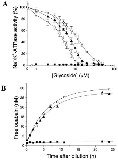
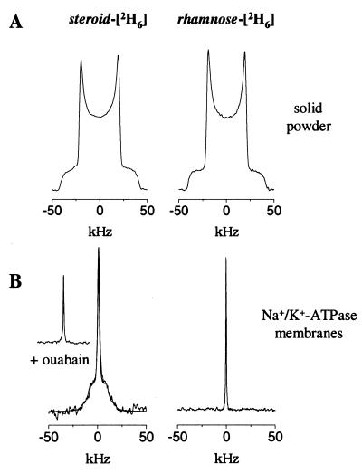
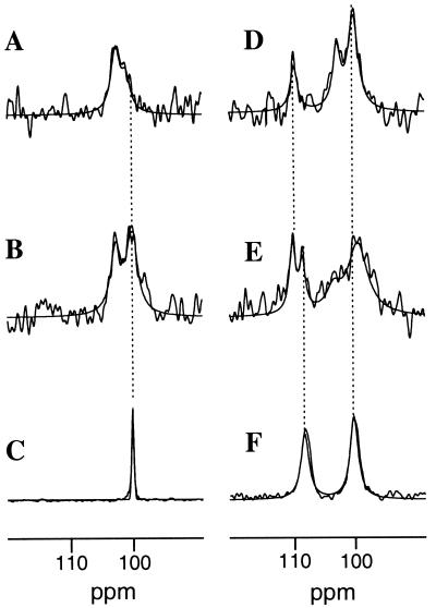
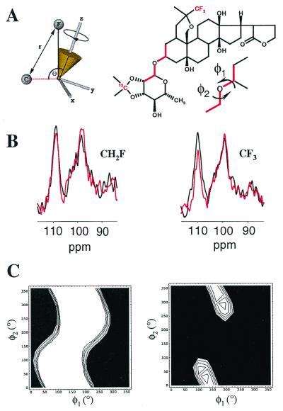
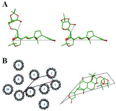
Several biologically active derivatives of the cardiotonic steroid ouabain have been made containing NMR isotopes ((13)C, (2)H, and (19)F) in the rhamnose sugar and steroid moieties, and examined at the digitalis receptor site of renal Na(+)/K(+)-ATPase by a combination of solid-state NMR methods. Deuterium NMR spectra of (2)H-labeled inhibitors revealed that the sugar group was only loosely associated with the binding site, whereas the steroid group was more constrained, probably because of hydrogen bonding to residues around the K(+)-channel region. Crosspolarization magic-angle spinning NMR showed that chemical shifts of inhibitors (13)C-labeled in the sugar group moved downfield by 0.5 ppm after binding to the digitalis site, suggesting that the sugar was close to aromatic side groups. A (19)F, (13)C- rotational-echo double-resonance NMR strategy was used to determine the structure of an inhibitor in the digitalis receptor site, and it showed that the ouabain derivatives adopt a conformation in which the sugar extends out of the plane of the steroid ring system. The combined structural and dynamic information favors a model for inhibition in which the ouabain analogues lie across the surface of the Na(+)/K(+)-ATPase alpha-subunit with the sugar group facing away from the surface of the membrane but free to move into contact with one or more aromatic residues.


Figures

Figure 1
Figure 1
Binding and activity data for ouabain and its acetonide derivatives in renal Na+/K+-ATPase membranes at 37°C. (A) Dose-response curve showing the ATPase activity remaining at the specified concentrations of ouabain (■), OMA (○), ODA (▴), and OFDA (◊) relative to the total activity in the absence of inhibitors. (B) The time course of release of [3H]ouabain from Na+/K+-ATPase after addition of 50 μM nonlabeled ouabain (□) or ODA (▴) was followed for up to 25 h. The stability of the enzyme-[3H]ouabain complex is also indicated (■).
Figure 2
Figure 2
Deuterium NMR spectra (recorded at 0°C) of [steroid-2H6]ODA (IV) and [rhamnose-2H6]ODA (V) in solid powder form (A) and when added to renal Na+/K+-ATPase membranes in molar equivalence to the digitalis sites (B). The spectrum after addition of an excess of ouabain is shown (Inset). The spectrum of IV in the membrane preparation (B Left) was simulated (using algorithms described in ref. 26) by superposition of a Lorenztian line shape and an axially symmetrical powder pattern with a residual quadrupole splitting of 17 kHz. The powder component represented >70% of the total signal.
Figure 3
Figure 3
Region of 13C CP-MAS NMR spectra of renal Na+/K+-ATPase membranes and 13C-labeled ouabain derivatives. Membrane spectra were recorded (at −50°C) in the absence of inhibitors (A) and after addition of [13C]OMA (II) in molar equivalence to the digitalis sites (B), and the spectrum was recorded of II in aqueous solution (C). Spectra also were recorded of membranes after the addition of [rhamnose-13C, steroid-13C]ODA (VI) in molar equivalence (D) and in a 2-fold excess (E) with respect to the digitalis site concentration, and of VI in aqueous solution (F). The dashed lines highlight the chemical shifts of II and VI in solution and II and VI in Na+/K+-ATPase membranes. Spectra resulted from the accumulation of 8192 scans with a repetition rate of 2 s.
Figure 4
Figure 4
Strategy for structure determination of VIII and IX at the digitalis site, from REDOR measurements of rhamnose-13C signal dephasing by the steroid-19F. (A) First, dephasing S/S0 was calculated for the time-dependent distance r between 13C and 19F (shown in red), for molecular conformations covering the entire conformational space of the rhamnose group. The calculations took into account the torsional angles φ1 and φ2, the coordinates defining the 19F rotational trajectory in a fixed molecular reference frame (x, y, z), angle θ between the C-F rotational axis and the 13C-C vector, the number of 19F atoms (three in compound VIII and one in compound IX), and the experimental NMR conditions. (B) Second, values of S/S0 were measured experimentally (at −50°C) from the resonance line at 110.4 ppm in the full-echo (black) and dephased-echo (red) spectra from either VIII (Left) or IX (Right) in the digitalis site. Spectra resulted from the accumulation of 54,000 scans. (C) Third, molecular conformations were determined by comparing the calculated S/S0 values with the experimental values. Torsional angles φ1 and φ2 giving the best fit of the calculated S/S0 to the experimental dephasing are identified from the light-colored regions of contour plots for VIII (Left) and IX (Right).
Figure 5
Figure 5
Structural features of cardiac glycosides and the digitalis site. (A) Representatives of the two groups of closely related structures of OFDA at the digitalis site determined by 13C,19F-REDOR NMR. Carbon atoms are shown in green, oxygen atoms in red, and hydroxyl groups are represented as spheres for clarity. (B) The 10 putative transmembrane regions of the Na+/K+-ATPase α subunit were fit to the electron density map of Ca2+-ATPase (6), showing in red the mutation sites conferring ouabain resistance to HeLa cells. One possible structure of OFDA is shown alongside the protein model to illustrate the comparative dimensions of the cardiac glycosides and the surface of the α subunit, and a possible docking orientation.

References

    1. Lingrel J B, Kuntzweiler T A. J Biol Chem. 1994;269:19659–19662. - PubMed
    1. Thomas R, Gray P, Andrews J. Adv Drug Res. 1990;19:311–562.
    1. Jewell E A, Shamraj O I, Lingrel J B. Acta Physiol Scand. 1992;146:161–169. - PubMed
    1. Bamberg E, Schoner W, editors. The Sodium Pump: Structure, Mechanism, Hormonal Control, and its Role in Disease. New York: Springer; 1994.
    1. Stokes D L, Auer M, Zhang P, Kuhlbrandt W. Curr Biol. 1999;9:672–679. - PubMed

Publication types

MeSH terms

Substances

LinkOut - more resources