. 2000 Dec 5;97(25):13631-6.
doi: 10.1073/pnas.250477397.

IFNalpha/beta promotes cell survival by activating NF-kappa B

Affiliations

IFNalpha/beta promotes cell survival by activating NF-kappa B

C H Yang et al. Proc Natl Acad Sci U S A. .

Abstract

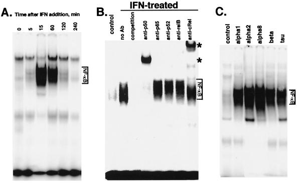
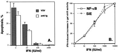
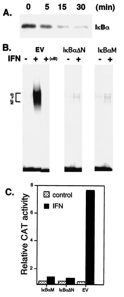
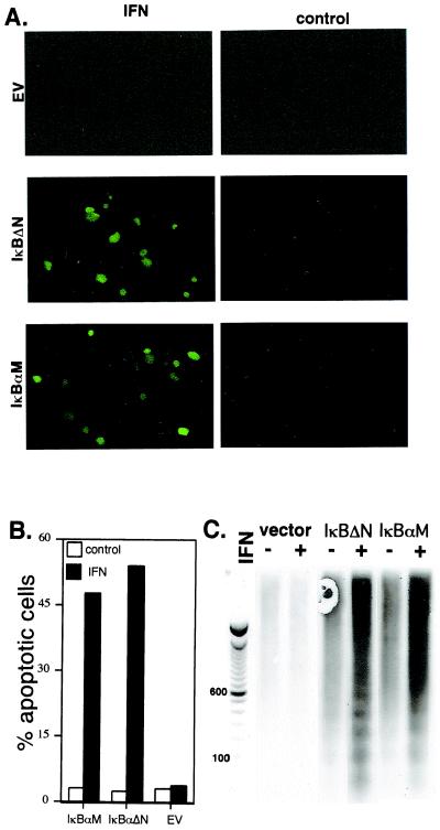
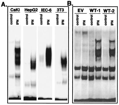
IFNs play critical roles in host defense by modulating the expression of various genes via signal transducer and activator of transcription factors. We show that IFNalpha/beta activates another important transcription factor, NF-kappaB. DNA-binding activity of NF-kappaB was induced by multiple type 1 IFNs and was promoted by IFN in a diverse group of human, monkey, rat, and murine cells. Human IFN promoted NF-kappaB activation in murine cells that express the human IFNalpha/beta receptor-1 signal-transducing chain of the type 1 IFN receptor. IFN promotes inhibitor of kappa B alpha (IkappaBalpha) serine phosphorylation and degradation, and stimulates NF-kappaB DNA-binding and transcriptional activity. Importantly, IFN promotes cell survival by protecting cells against a variety of proapoptotic stimuli, such as virus infection and antibody-mediated crosslinking. Expression of superrepressor forms of IkappaBalpha, besides inhibiting IFN-mediated NF-kappaB activation and IkappaBalpha degradation, also enhanced apoptotic cell death in IFN-treated cells. We conclude that NF-kappaB activation by IFNalpha/beta is integrated into a signaling pathway through the IFNalpha/beta receptor-1 chain of the type 1 IFN receptor that promotes cell survival in apposition to various apoptotic stimuli.


Figures

Figure 1
Figure 1
IFN induces NF-κB activation. (A) EMSA on nuclear extracts from Daudi cells treated with IFNCon1 (5,000 units/ml) for varying times. (B) Nuclear extracts from Daudi cells treated with IFNCon1 (5,000 units/ml; 30 min) were subjected to EMSA in the absence or presence of a 50-fold excess of unlabeled κB oligonucleotide probe (competition). *, Positions of supershifted complexes. (C) EMSA on nuclear extracts from Daudi cells treated with various type 1 IFNs (5,000 units/ml) for 30 min.
Figure 2
Figure 2
IFN promotes cell survival. (A) Daudi cells were pretreated overnight with various concentrations of IFNCon1 and analyzed for apoptosis by TUNEL assays at 1 day after either infection by VSV or anti-Ig treatment. The data shown in the graph are the average of two experiments performed in duplicate and are expressed in terms of apoptosis induced by VSV or anti-Ig. (B) Dose-dependent NF-κB activation and sis-inducible element gel shift activity by IFN. Nuclear extracts from Daudi cells treated with varying concentrations of IFNCon1 (0–5,000 units/ml) for 30 min were incubated with a 32P-endlabeled promoter probe for the consensus κB site or the high affinity sis-inducible element (SIE) from the c-fos gene (5′-AGCTTCATTTCCCGTAATCCCTAAAGCT-3′). EMSA results were quantitated on a PhosphorImager (Molecular Dynamics) by using quantity one software (Bio-Rad). The results of three experiments were averaged (SEM < 20%) and expressed relative to the increased gel shift activity observed at an IFN concentration of 10,000 units/ml.
Figure 3
Figure 3
The role of IκBα in NF-κB activation by IFNα/β. (A) Cell lysates from Daudi cells treated with IFNCon1 (5,000 units/ml) were resolved by SDS/PAGE, blotted onto poly(vinylidene difluoride) membranes, probed with anti-IκBα, and visualized by enhanced chemiluminescence. (B) EMSA with a 32P-labeled κB probe on nuclear extracts from control and IFN-treated Daudi cells transiently transfected for 48 h with IκBαM, IκBαΔN, or with empty vector (EV). Binding to the κB probe was blocked by a 50-fold excess of unlabeled probe (+ κB). (C) NF-κB-dependent reporter gene activity in IFN-treated COS cells transiently cotransfected with the pUX-CAT 3XHLAκB construct and IκBαM, IκBαΔN, or empty vector. At 2 days after transfection, cells were treated in the presence or absence of IFN for 30 min, lysed, and assayed for CAT activity as determined by phosphorimaging. Data shown are the average of three experiments (SEM < 15%), expressed relative to CAT activity in cells transfected with empty vector.
Figure 4
Figure 4
The role of IκBα in IFNα/β promoted cell survival. IFN-treated (1,000 units/ml; 24 h) Daudi cells transiently transfected for 48 h with IκBαM, IκBαΔN, or empty vector were analyzed for apoptosis by TUNEL assays (A and B) or apoptotic DNA by a chemiluminescent assay with a DNA ladder provided for reference (C). Apoptotic cells are fluorescent and thus appear green in the photomicrograph. The TUNEL data (B) represent the mean of three independent experiments in which at least 500 cells were scored for each variable (SEM < 10%).
Figure 5
Figure 5
IFN induces NF-κB activation in diverse cell types. (A) EMSA on nuclear extracts from IFN-treated (5,000 units/ml rat IFNβ for 30 min) murine 3T3 fibroblasts and rat intestinal IEC-6 cells, or IFN-treated (5,000 units/ml IFNCon1 for 30 min) human CaKi and HepG2 cells. (B) Murine L929 cell transfectants expressing empty vector (EV) or two independent wild-type huIFNAR-1 (WT) transfectants (29) were treated with IFNCon1 (5,000 units/ml) for 30 min before gel shift analysis with a NF-κB probe.

References

    1. Branca A A, Faltynek C R, D'Alessandro S B, Baglioni C. J Biol Chem. 1982;257:13291–13296. - PubMed
    1. Uze G, Lutfalla G, Gresser I. Cell. 1990;60:225–234. - PubMed
    1. Novick D, Cohen B, Rubinstein M. Cell. 1994;77:391–400. - PubMed
    1. Domanski P, Witte M, Kellum M, Rubinstein M, Hackett R, Pitha P, Colamonici O R. J Biol Chem. 1995;270:21606–21611. - PubMed
    1. Friedman R L, Stark G R. Nature (London) 1985;314:637–639. - PubMed

Publication types

LinkOut - more resources