. 2000 Dec 5;97(25):13796-800.
doi: 10.1073/pnas.250492697.

Haploinsufficiency of Snf5 (integrase interactor 1) predisposes to malignant rhabdoid tumors in mice

Affiliations

Haploinsufficiency of Snf5 (integrase interactor 1) predisposes to malignant rhabdoid tumors in mice

C W Roberts et al. Proc Natl Acad Sci U S A. .

Abstract

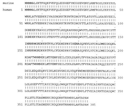
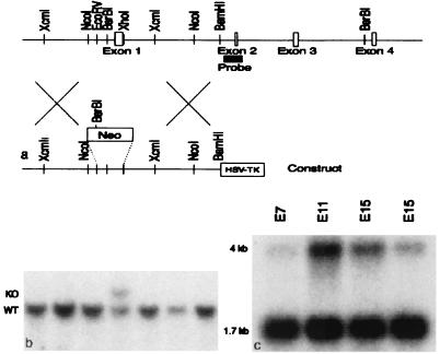
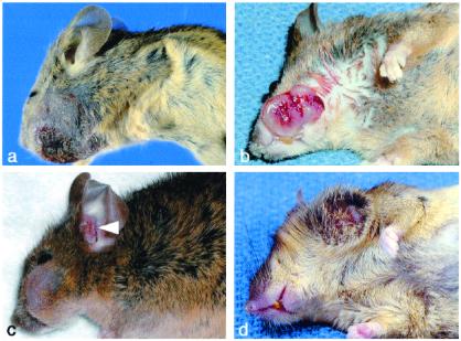
Malignant rhabdoid tumor (MRT) is an aggressive, highly lethal cancer of young children. Tumors occur in various locations, including kidney, brain, and soft tissues. Despite intensive therapy, 80% of affected children die, often within 1 year of diagnosis. The majority of MRT samples and cell lines have sustained biallelic inactivating mutations of the hSNF5 (integrase interactor 1) gene, suggesting that hSNF5 may act as a tumor suppressor. We sought to examine the role of Snf5 in development and cancer in a murine model. Here we report that Snf5 is widely expressed during embryogenesis with focal areas of high-level expression in the mandibular portion of the first branchial arch and central nervous system. Homozygous knockout of Snf5 results in embryonic lethality by embryonic day 7, whereas heterozygous mice are born at the expected frequency and appear normal. However, beginning as early as 5 weeks of age, heterozygous mice develop tumors consistent with MRT. The majority of tumors arise in soft tissues derived from the first branchial arch. Our findings constitute persuasive genetic evidence that Snf5, a core member of the Swi/Snf chromatin-remodeling complex, functions as a tumor suppressor gene, and, moreover, Snf5 heterozygotes provide a murine model of this lethal pediatric cancer.


Figures

Figure 1
Figure 1
Alignment of murine and human Snf5.
Figure 2
Figure 2
Snf5 expression during development. Wild-type embryos were harvested at E8 (a), E9.5 (b), and E11.5 (c and d). In situ hybridization was carried out with antisense (a, b, and d) or sense (c) probes to Snf5. Expression is detected throughout the embryos but is particularly intense in the headfolds (black arrows), neural folds (black arrowheads), first branchial arch (white arrow), and hindlimb bud (white arrowheads).
Figure 3
Figure 3
Homologous recombination knockout construct and embryonic expression of Snf5. (a) The murine Snf5 genomic locus and targeting construct. (b) Southern blot of embryonic stem cell DNA cut with BsrBI and XhoI and hybridized with the probe. The wild-type band is 11.3 kb, and the targeted band is 12.5 kb. (c) Northern blot of murine whole-embryo RNA hybridized with a probe covering the entire ORF of the cDNA of murine Snf5. Transcripts of 1.7 and 4 kb are detected.
Figure 4
Figure 4
Gross appearance of tumors. (a) Mouse 32. (b) Mouse 130. (c) Mouse 5. Two tumors are present: one on the lateral face and a metachronous/metastatic tumor in the external ear canal (arrowhead). (d) Mouse 53.
Figure 5
Figure 5
Microscopic appearance of tumors. (a) Low-power view demonstrating areas of necrosis (N), hemorrhage (H), and epithelial invasion (E) (×40). (b and c) Medium-power and high-power views revealing classic rhabdoid cells (arrows) with large eccentric nuclei, prominent nucleoli, and a prominent eosinophilic cytoplasm (×400, ×1,000). (d) Lymph node with subcapsular metastasis (×200). (e) Lung with metastatic tumor (×200). (f) EM of rhabdoid cell demonstrating large cytoplasm containing whorls of intermediate filaments. (g and h) Corresponding sections demonstrating H&E and immunohistochemical staining for Snf5 in tumor infiltrating skeletal muscle. Entrapped eosinophilic muscle fibers (S) are positive for Snf5, whereas the tumor cells (T) are negative (×200).

References

    1. Kingston R E, Narlikar G J. Genes Dev. 1999;13:2339–2352. - PubMed
    1. Peterson C L, Dingwall A, Scott M P. Proc Natl Acad Sci USA. 1994;91:2905–2908. - PMC - PubMed
    1. Cote J, Quinn J, Workman J L, Peterson C L. Science. 1994;265:53–60. - PubMed
    1. Cairns B R, Kim Y-J, Sayre M H, Laurent B C, Kornberg R D. Proc Natl Acad Sci USA. 1994;91:1950–1954. - PMC - PubMed
    1. Sudarsanam P, Winston F. Trends Genet. 2000;16:345–351. - PubMed

Publication types