Review
. 2000;9(1-2):77-91.
doi: 10.3727/000000001783992696.

Chemical approaches to control gene expression

Affiliations
Review

Chemical approaches to control gene expression

J M Gottesfeld et al. Gene Expr. 2000.

Abstract

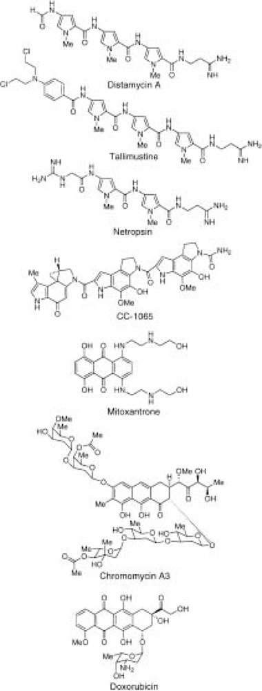
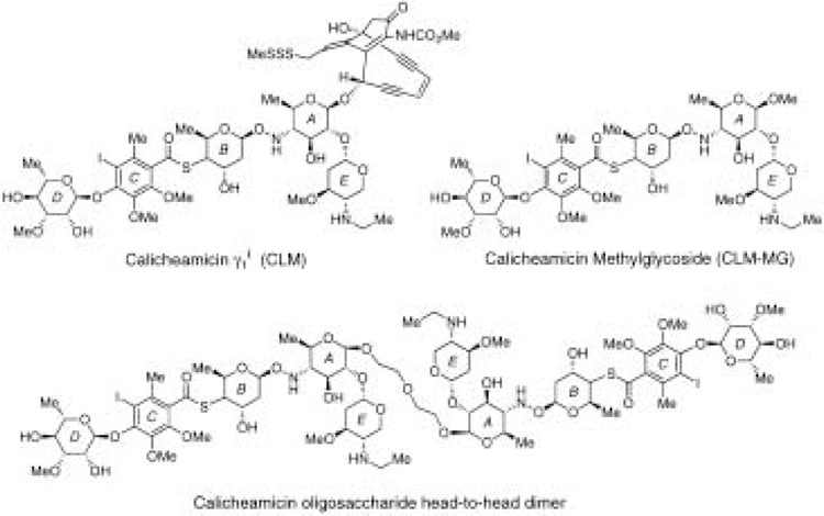
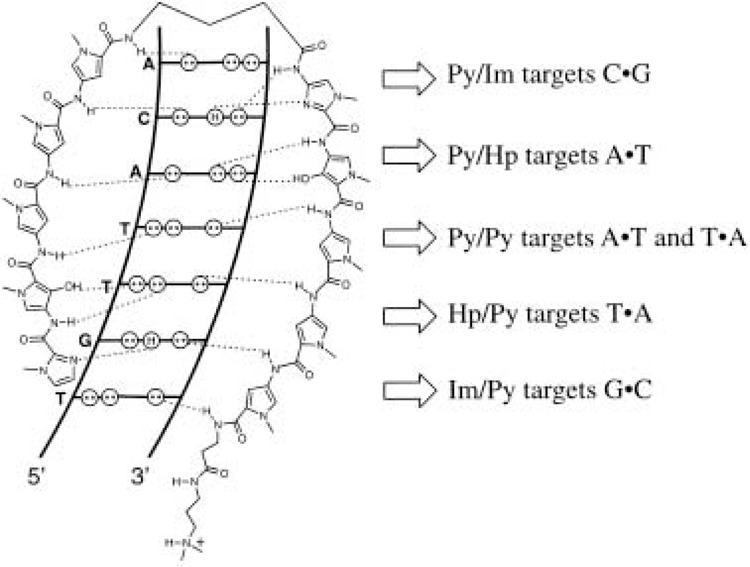
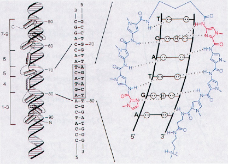
A current goal in molecular medicine is the development of new strategies to interfere with gene expression in living cells in the hope that novel therapies for human disease will result from these efforts. This review focuses on small-molecule or chemical approaches to manipulate gene expression by modulating either transcription of messenger RNA-coding genes or protein translation. The molecules under study include natural products, designed ligands, and compounds identified through functional screens of combinatorial libraries. The cellular targets for these molecules include DNA, messenger RNA, and the protein components of the transcription, RNA processing, and translational machinery. Studies with model systems have shown promise in the inhibition of both cellular and viral gene transcription and mRNA utilization. Moreover, strategies for both repression and activation of gene transcription have been described. These studies offer promise for treatment of diseases of pathogenic (viral, bacterial, etc.) and cellular origin (cancer, genetic diseases, etc.).


Figures

Figure 1
Figure 1
Structures of small molecules that bind DNA.
Figure 2
Figure 2
Structures of calicheamicin γ1 I (CLM), the methylglycoside of the carbohydrate moiety of CLM (CLM-MG), and the head-to-head dimer of the CLM oligosaccharide.
Figure 3
Figure 3
Binding model for the complex formed between ImHpPyPyPy-y-ImHpPyPyPy-P-Dp and a 5′-TGTTACA-3′ sequence. Circles with dots represent the lone pairs of N(3) of purines and O(2) of pyrimidines, and circles containing an H represent the 2-amino group of guanine. Hydrogen bonds are shown as dashed lines.
Figure 4
Figure 4
Left: model of the nine-zinc finger protein TFIIIA with the 5S RNA gene internal control region (ICR). Middle: sequence of the ICR recognized by zinc finger 4 in the minor groove. Right: complex of a polyamide with its target site, 5′-AGTACT-3′. Circles with dots represent the lone pairs of N(3) of purines and O(2) of pyrimidines, and circles containing an H represent the 2-amino group of guanine. Hydrogen bonds are shown as dashed lines.
Figure 5
Figure 5
Schematic of the HIV-1 enhancer and promoter (nucleotide positions −199 to +1) showing binding sites for polyamides and the transcription factors upstream stimulatory factor (USF), Ets-1, LEF-1, NF-kB, Sp1, and TFIID (TBP). For polyamide binding models: shaded and unshaded circles, Im and Py rings, respectively; curved lines, γ-aminobutyric acid (γ); diamonds, β-alanine (β); and Dp, dimethylamino-propylamide. (a) A polyamide binds to sequences adjacent to the TATA box element of the HIV-1 promoter and inhibits TBP binding. (b) Polyamides bind adjacent to the binding sites for LEF-1 and Ets-1 and inhibit binding of these transcription factors.
Figure 6
Figure 6
Sequence of the human cytomegalovirus major immediate early promoter region from position —34 to position +8. The TATA box and the repressor binding site (located at —14 to +1 relative to the start site of transcription) are boxed. (a) Without inhibition, repressor binds and blocks transcription. (b) Polyamide binding inhibits repressor binding, and transcription occurs. The polyamide is schematically represented at its DNA-binding site. The black and white circles represent imidazole and pyrrole rings, respectively; the hairpin junction (curved line) is formed with γ-aminobutyric acid, and the diamond represents β-alanine.
Figure 7
Figure 7
A schematic model of Arg-Pro-Arg (RPR) polyamides targeted to the major groove transcription factor GCN4. (A) The a-helical GCN4 dimer (yellow) is shown binding to adjacent major grooves. The Arg-Pro-Arg-hairpin polyamide is shown as red, blue, and green balls, which represent imidazole, pyrrole, and Arg-Pro-Arg amino acids, respectively. The blue diamond represents β-alanine. γ-Aminobutyric acid is designated as a curved line. (B) The contacts between one GCN4 monomer and the major groove of one half site of 5′-CTGACTAAT-3′ are depicted. Circles with two dots represent the lone pairs of the N7 of purines, the O4 or thymine and the O6 of guanine. Circles containing an H represent the N6 and N4 hydrogens of the exocyclic amines of adenine and cytosine, respectively. The C5 methyl group of thymine is depicted as a circle with CH3 inside. Protein side chains that make hydrogen bonds or van der Waals contacts to the bases are shown in purple and connected to the DNA via a dotted line. Green and purple plus signs represent protein residues that electrostatically contact the phosphate backbone. The residues that are predicted to be disrupted by an Arg-Pro-Arg polyamide are shown in green. (C) The hydrogen-bonding model of the eight-ring hairpin polyamide ImPyPyPy-y-PyPyPyPy-P-RPR bound to the minor groove of 5′-TGTTAT-3′. Circles with two dots represent the lone pairs of N3 of purines and O2 of pyrimidines. Circles containing an H represent the N2 hydrogens of guanines. Putative hydrogen bonds are illustrated by dotted lines. Py and Im rings are represented as blue and red rings, respectively. The Arg-Pro-Arg moiety is green. (D) The model of the polyamide binding its target site (bold) adjacent to the GCN4 binding site (brackets). Polyamide residues are as in (A).
Figure 8
Figure 8
A synthetic activator consisting of an activation domain (AD), a dimerization or linker domain (LD), and a DNA-binding domain (DBD) complexed with the cognate palindromic DNA site of the hairpin polyamide-peptide conjugate. The black and white circles represent imidazole and pyrrole rings, respectively, and the hairpin junction (curved line) is formed with γ-aminobutyric acid.
Figure 9
Figure 9
Structures of metallointercalators that bind DNA in the major groove.
Figure 10
Figure 10
Structures of two xanthene tetraureas active in binding to DNA.

References

    1. Beerli R. R.; Segal D. J.; Dreier B.; Barbas C. F. 3rd. Toward controlling gene expression at will: Specific regulation of the erbB-2/HER-2 promoter by using polydactyl zinc finger proteins constructed from modular building blocks. Proc. Natl. Acad. Sci. USA 95:14628–14633; 1998. - PMC - PubMed
    1. Bellorini M.; Moncollin V.; D’Incalci M.; Mongelli N.; Mantovani R. Distamycin A and tallimustine inhibit TBP binding and basal in vitro transcription. Nucleic Acids Res. 23:1657–1673; 1995. - PMC - PubMed
    1. Bianchi N.; Passadore M.; Rutigliano C.; Feriotto G.; Mischiati C.; Gambari R. Targeting of the Sp1 binding sites of HIV-1 long terminal repeat with chro-momycin. Disruption of nuclear factor.DNA complexes and inhibition of in vitro transcription. Biochem. Pharmacol. 52:1489–1498; 1996. - PubMed
    1. Bohjanen P. R.; Liu Y.; Garcia-Blanco M. A. TAR RNA decoys inhibit tat-activated HIV-1 transcription after preinitiation complex formation. Nucleic Acids Res. 25:4481–4486; 1997. - PMC - PubMed
    1. Breaker R. R. Catalytic DNA: In training and seeking employment. Nat. Biotechnol. 17:422–423; 1999. - PubMed

Publication types

LinkOut - more resources