Review
. 2000 Dec 1;290(5497):1717-21.
doi: 10.1126/science.290.5497.1717.

Autophagy as a regulated pathway of cellular degradation

Affiliations
Review

Autophagy as a regulated pathway of cellular degradation

D J Klionsky et al. Science. .

Abstract

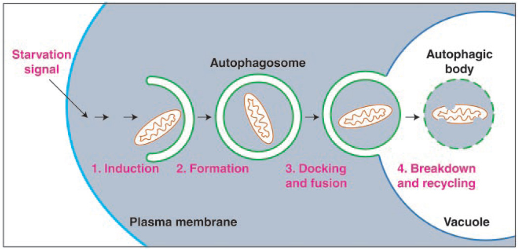
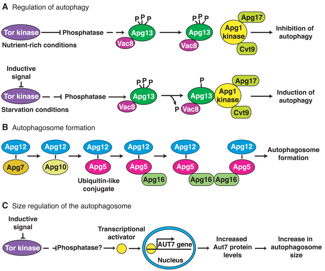
Macroautophagy is a dynamic process involving the rearrangement of subcellular membranes to sequester cytoplasm and organelles for delivery to the lysosome or vacuole where the sequestered cargo is degraded and recycled. This process takes place in all eukaryotic cells. It is highly regulated through the action of various kinases, phosphatases, and guanosine triphosphatases (GTPases). The core protein machinery that is necessary to drive formation and consumption of intermediates in the macroautophagy pathway includes a ubiquitin-like protein conjugation system and a protein complex that directs membrane docking and fusion at the lysosome or vacuole. Macroautophagy plays an important role in developmental processes, human disease, and cellular response to nutrient deprivation.


Figures

Fig. 1
Fig. 1
Schematic model of macroautophagy in yeast. A signal transduction event regulated by the Tor kinase leads to the following: (1) The induction of autophagy. (2) Membrane from an unknown source sequesters cytosol and/or organelles (a mitochondrion is depicted) resulting in the formation of a double-membrane vesicle (300 to 900 nm) termed an autophagosome. (3) On completion, the autophagosome docks with the lysosome or vacuole. Fusion of the autophagosome outer membrane with the vacuole releases the inner vesicle into the vacuole lumen. The inner vesicle is termed an autophagic body. (4) Breakdown within the vacuole allows recycling of the degraded autophagic body and its hydrolyzed cargo (amino acids, fatty acids, sugars, and nucleotides). The morphology of macroautophagy in mammalian cells is similar to that shown; however, in mammalian cells autophagy can be induced by environmental cues other than starvation.
Fig. 2
Fig. 2
Molecular genetics of macroautophagy in yeast. (A) The Tor kinase exerts a negative regulatory effect on autophagy when cells are growing under nutrient-rich conditions. When starvation occurs, the Tor kinase is inactivated, and the negative regulation is relieved resulting in induction of autophagy. Most of the proteins required for autophagy are constitutively expressed and are used for biosynthetic import through the cytoplasm to vacuole targeting pathway under these conditions. The downstream effectors of Tor are likely to include phosphatases and kinases that modulate the phosphorylation state of Apg13. An inductive signal such as carbon or nitrogen starvation inactivates Tor and results in partial dephosphorylation of Apg13. This form of Apg13 associates more tightly with the Apg1 kinase and stimulates its activity. The function of Apg1 kinase is required for autophagosome formation. (B) The Apg7 (E1-like) and Apg10 proteins form thioester intermediates through a COOH-terminal glycine of Apg12. Apg12 is ultimately conjugated to Apg5 through an internal lysine residue in Apg5 in a process that is similar to ubiquitination. Apg16 binds the conjugated Apg5 protein noncovalently and dimerizes to form a complex that is required for formation and completion of the autophagosome. (C) Under nutrient-rich conditions, the Tor kinase negatively regulates the expression of the AUT7 gene resulting in basal levels of Aut7 synthesis. Under these conditions the Cvt pathway is operative and 150-nm Cvt vesicles are formed. Inhibition of Tor by transduction of an environmental signal or after treatment with rapamycin allows the activation of a presumed transcriptional activator protein that increases expression of AUT7. The resulting increase in Aut7 levels allows an expansion in the size of the autophagosome from 150 nm to a range of 300 to 900 nm.

References

    1. Mortimore GE, Miotto G, Venerando R, Kadowaki M. Subcell. Biochem. 1996;27:93. - PubMed
    1. Vittorini S, et al. J. Gerontol. A Biol. Sci. Med. Sci. 1999;54:B318. - PubMed
    1. Dunn WA., Jr Trends Cell Biol. 1994;4:139. - PubMed
    1. Cuervo AM, Dice JF. J. Mol. Med. 1998;76:6. - PubMed
    1. Kim J, Klionsky DJ. Annu. Rev. Biochem. 2000;69:303. - PubMed

Publication types