. 2000 Dec 1;19(23):6331-40.
doi: 10.1093/emboj/19.23.6331.

The interaction of titin and alpha-actinin is controlled by a phospholipid-regulated intramolecular pseudoligand mechanism

Affiliations

The interaction of titin and alpha-actinin is controlled by a phospholipid-regulated intramolecular pseudoligand mechanism

P Young et al. EMBO J. .

Abstract

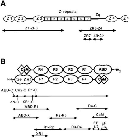
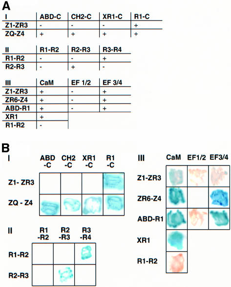
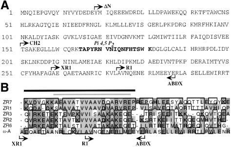
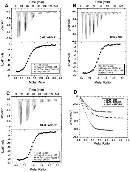
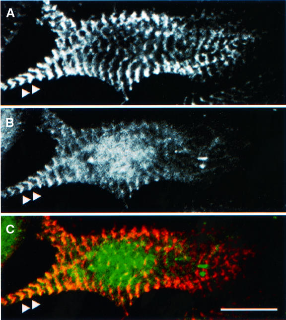
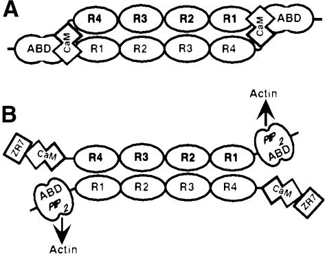
The assembly of stable cytoskeletal structures from dynamically recycled molecules requires developmental and spatial regulation of protein interactions. In muscle, titin acts as a molecular ruler organizing the actin cytoskeleton via interactions with many sarcomeric proteins, including the crosslinking protein alpha-actinin. An interaction between the C-terminal domain of alpha-actinin and titin Z-repeat motifs targets alpha-actinin to the Z-disk. Here we investigate the cellular regulation of this interaction. alpha-actinin is a rod shaped head-to-tail homodimer. In contrast to C-terminal fragments, full-length alpha-actinin does not bind Z-repeats. We identify a 30-residue Z-repeat homologous sequence between the actin-binding and rod regions of alpha-actinin that binds the C-terminal domain with nanomolar affinity. Thus, Z-repeat binding is prevented by this 'pseudoligand' interaction between the subunits of the alpha-actinin dimer. This autoinhibition is relieved upon binding of the Z-disk lipid phosphatidylinositol-bisphosphate to the actin-binding domain. We suggest that this novel mechanism is relevant to control the site-specific interactions of alpha-actinin during sarcomere assembly and turnover. The intramolecular contacts defined here also constrain a structural model for intrasterical regulation of all alpha-actinin isoforms.


Figures

None
Fig. 1. (A) Domain structure of the Z-disk region of titin. Z1, Z2, Z3 and Z4 = immunoglobulin-like domains; ZR1–ZR7 = Z-repeats; Zq = non-modular region. Only ∼100 kDa of the 3 MDa titin molecule are shown. (B) Schematic diagram of the α-actinin anti-parallel dimer showing the domain structure of the molecule. The N-terminal actin-binding domain is composed of two calponin homology domains. The central rod region consists of four spectrin-like repeats. A CaM-like domain is located at the C-terminus of the molecule. ABD = actin binding domain; CH = calponin homology domain; R1, R2, R3 and R4 = spectrin-like repeats; CaM = calmodulin-like domain. Shown below each molecule are the constructs used in this study. See Materials and methods for the amino acid sequence of these constructs. The nomenclature indicates the most N- and C-terminal domains in a given construct. ‘-C’, constructs that contain the C-terminus of the molecule.
None
Fig. 2. Yeast two-hybrid analysis of the intersubunit and titin interactions of the α-actinin dimer. α-actinin-2 and titin constructs were cloned into the pLexA or pGAD10 vectors and cotransformed into the reporter yeast strain. Interactions between constructs, as shown by expression of the His3 and β-galactosidase reporter genes, was monitored by scoring growth on plates lacking histidine (A) or in colorimetric filter based assays using X-gal as substrate (B). pLexA constructs are shown on the left and pGAD10 constructs on the top of each grid/table. (I) Interactions between titin constructs (Z1-ZR3 and ZQ-Z4) and N-terminally truncated α–actinin molecules. See Figure 1 for details of the nomenclature of the constructs. (II) Interactions between pairs of repeats from the α-actinin rod. (III) Interactions of the α-actinin CaM-like domain with titin constructs containing Z-repeats and with the α-actinin ABD-R1 construct. Division of the CaM domain into two halves (EF1/2 and EF3/4) shows that both binding activities are mediated by EF-hands 3/4. An R1 construct, XR1 extended at the N-terminus by 15 residues, is sufficient to mediate the interaction with the CaM domain.
None
Fig. 3. (A) Sequence of the first 300 amino acids of skeletal muscle α-actinin 2A. N-termini and C-termini of the various constructs used are indicated. Highlighted in bold is the PI4,5P2 binding site as identified by Fukami et al., (1996). (B) Sequence alignment of five of the titin Z-repeats (numbered according to Sorimachi et al., 1997) with a region of α-actinin (α-A) that spans the C-terminal end of the actin-binding domain and the beginning of the first repeat of the rod. Homologous residues are boxed, non-polar residues are shaded light grey, charged residues are shaded dark grey. Note the hydrophobic cluster of residues in the first half of the alignment, which is the hallmark of the Z-repeats. α-actinin residues 259–307 can be aligned with the Z-repeats and share a similar hydrophobic region as well as some other conserved residues. Positions of the N- and C-termini of various constructs are indicated. The dark bar indicates the α-actinin binding site within ZR7 defined by Ohtsuka et al. (1997b). The light bar indicates the region of ZR7 that is helical when in complex with the α-actinin EF3/4 region (Atkinson et al., 2000).
None
Fig. 4. Proteins used in this study. Recombinant proteins were expressed in E.coli as described and analysed by SDS–PAGE with Coomassie Blue staining. Calculated molecular weights (in kDa) are shown in brackets. Lanes 1–9: α-actinin constructs (2A isoform unless stated otherwise). Lane 1: ABD-C (104); lane 2: ΔN-C (102); lane 3: CH2-C (87); lane 4: XR1-C (74); lane 5: R1-C (73); lane 6: α-actinin-1A R1-C (73); lane 7: R4-C(31); lane 8: ABD-R1(46); lane 9: CaM-like domain (17). Lanes 10–13: titin constructs. Lane 10: GST–ZR7 (34); lane 11: GST (29); lane 12: GST–Zq-Δ6 (43); lane 13: ZR7 (6). M, molecular weight markers (sizes shown in kDa). Note that the GST–ZR7 fusion protein shows slight degradation. The lower band is GST that has lost the ZR7 fragment during purification.
None
Fig. 5. Binding of α-actinin constructs to GST–ZR7. Proteins were mixed and bound to GST affinity beads as described. Proteins that remained bound after washing were eluted with loading buffer and visualized by SDS–PAGE with Coomassie Blue staining. Lane 1: α-actinin-2 ABD-C/GST–ZR7; lane 2: α-actinin-2 ΔN-C/GST–ZR7; lane 3: α-actinin-2 CH2-C/GST–ZR7; lane 4: α-actinin-2 R1ext-C/GST–ZR7; lane 5: α-actinin-2 R1-C/GST–ZR7; lane 6: α-actinin-2 R1-C/GST; lane 7: α-actinin-1 R1-C/GST-ZR7; lane 8: α-actinin-1A R1-C/GST. M, molecular weight marker. Note, a faint band of 60 kDa in the GST control lanes probably represents some dimeric GST. Bound α-actinin R1-C is indicated by the arrowhead.
None
Fig. 6. (A) Competitive binding of α-actinin-2 ABD-R1 and titin ZR7 to the α-actinin-2 CaM-like domain (CaM). Proteins were mixed and bound to Ni–NTA agarose as described and the fractions analysed by SDS–PAGE with Coomassie Blue staining. The flow-through and wash fractions were mixed and loaded in lanes 1–4. Bound proteins were eluted with loading buffer and run in lanes 5–8. Lanes 1 and 5: His6-A BDX/CaM; lanes 2 and 6: His6-ABD-R1/CaM; lanes 3 and 7: His6-ABD-R1/CaM/20 µM ZR7; lanes 4 and 8: His6-ABD-R1/CaM/100 µM ZR7. The arrowhead indicates the bound CaM-like domain. ZR7 stains poorly by Coomassie and is only visible at the highest concentration used (lane 4). (B) An α-actinin peptide comprising amino acids 259–287 inhibits binding of ABD-R1 to the CaM-like domain. The α-actinin His6-ABD-R1 and CaM constructs are mixed with increasing concentrations of peptide and bound to Ni–NTA agarose as in (A). After washing, bound proteins are eluted with loading buffer. Lanes 1–4: flow-through and wash fractions mixed; lanes 5–8: eluate fractions. Lanes 1 and 5: no peptide; lanes 2 and 6: 30 µM peptide; lanes 3 and 7: 150 µM peptide; lanes 4 and 8: 600 µM peptide. The arrowheads indicate the bound CaM-like domain.
None
Fig. 7. Inter-subunit and titin interactions of α-actinin demonstrated by isothermal calorimetry. The α-actinin CaM-like domain (CaM) or R4-C was titrated against ABD-R1 or titin ZR7 by injections of equal volume at regular time intervals. (A) 150 µM CaM and 10 µM ABD-R1; (B) 90 µM CaM and 9 µM ZR7; (C) 215 µM R4-C and 10 µM ABD-R1. The top graph in each case shows the raw data; the heat of the binding reaction is monitored as the differential energy (µcal/s) required to maintain the experimental cell at the same temperature (26°C) as the reference cell. The lower graph shows the processed data after normalization per mol of injectant, integration with respect to time and subtraction of reference data. The solid line is the calculated best fitting curve to this data obtained by optimization of the fitting parameters stoichiometry (n), enthalpy (ΔH) and association constant (Kb) using the least squares minimization routines provided in the Origin software. The values of these parameters obtained for each interaction are shown. (D) The integral mode of the data in A–C; integrated enthalphies are plotted against molar ratios of the respective proteins.
None
Fig. 8. (A) Effects of phospholipids on binding of full-length α-actinin to titin constructs, GST–ZR7 and GST–Zq-Δ6. Proteins were mixed and bound to GST affinity beads as described. Proteins that remained bound after washing were eluted with loading buffer and analysed by SDS–PAGE. Phospholipids were added to protein mixture as micelles in 0.5% Triton. Lane 1: α-actinin-2 ABD-C/GST–ZR7, control with 0.5% Triton; lanes 2, 3 and 4: α-actinin-2 ABD-C/GST–ZR7, 5, 10 and 25 µg/ml phosphatidylserine, respectively; lanes 5, 6 and 7: α-actinin-2 ABD-C/GST–ZR7, 5, 10 and 25 µg/ml phosphatidylinositol bisphosphate (PI4,5P2); lane 8: α-actinin-2 ABD-C/GST–ZR7, 25 µg/ml phosphatidylcholine; lane 9: native α-actinin-1A/GST–ZR7, no phospholipid; lane 10: native α-actinin-1A/GST–ZR7, 25 µg/ml PI4,5P2; lane 11: α-actinin-2 ABD-C/GST–ZR7, 600 µM d-myo-inositol 1,4,5-triphosphate; lane 12: α-actinin-2 ABD-C/GST–ZR7, 600 µM l-α-glycerophospho-d-myo-inositol 4,5-bisphosphate; lane 13: α-actinin-2 ABD-C/GST–Zq-Δ6, no phospholipid. (B) Effects of PI4,5P2 on binding of truncated α-actinin-2 constructs to GST–ZR7. PI4,5P2, as micelles in 0.5% Triton, was added at 25 µg/ml in lanes 1–3. Triton micelles alone were used as control in lanes 4–6. Lanes 1 and 4: α-actinin-2 ABD-C; lanes 2 and 5: α-actinin-2 CH2-C; lanes 3 and 6: α-actinin-2 XR1-C.
None
Fig. 9. Localization of PiP-4P-5K in neonatal rat cardiomyocytes. Endogenous proteins were visualized by indirect immunofluorescence using titin T12 to label the Z-disks (A) and a polyclonal antibody labelling PiP-4P-5K (B). Note that PiP-4P-5K is arranged in a striated sarcomeric pattern and the overlay with titin T12 (C) demonstrates its localization close to the Z-band (arrowheads). Note that the sarcomeric association is most pronounced in peripheral regions of the cell and that there is also a significant pool of apparently cytosolic protein. Scale bar, 10 µm.
None
Fig. 10. Model for the regulation of titin Z-repeat binding in α-actinin by phospholipids. (A) The closed or inactive state of the molecule when the EF3/4 region of the CaM-like domain interacts with a region between the ABD and R1 of the opposite subunit. Thus, binding of titin Z-repeats to EF3/4 is prevented. (B) Binding of PI4,5P2 to the ABD induces a conformational change that switches on titin binding by making the EF3/4 region available for binding to Z-repeats (ZR7). PI4,5P2 binding also increases the actin-binding properties of α-actinin (Fukami et al., 1992).

References

    1. Astier C., Raynaud,F., Lebart,M.C., Roustan,C. and Benyamin,Y. (1998) Binding of a native titin fragment to actin is regulated by PIP2. FEBS Lett., 429, 95–98. - PubMed
    1. Atkinson R.A., Joseph,C., Dal Piaz,F., Birolo,L., Stier,G., Pucci,P. and Pastore,A. (2000) Binding of α-actinin to titin: implications for Z-disk assembly. Biochemistry, 39, 5255–5264. - PubMed
    1. Ausubel F.M., Brent,R., Kingston,R.E., Moore,D.D., Seidman,J.G., Smith,J.A. and Struhl,K. (1987) Current Protocols in Molecular Biology. Wiley and Sons, Inc., New York, NY.
    1. Beckerle M.C. (1997) Zyxin: zinc fingers at sites of cell adhesion. BioEssays, 19, 949–957. - PubMed
    1. Blanchard A., Ohanian,V. and Critchley,D. (1989) The structure and function of α-actinin. J. Muscle Res. Cell Motil., 10, 280–289. - PubMed

Publication types

MeSH terms