. 2000 Dec 1;19(23):6371-81.
doi: 10.1093/emboj/19.23.6371.

Adenylate kinase 1 gene deletion disrupts muscle energetic economy despite metabolic rearrangement

Affiliations

Adenylate kinase 1 gene deletion disrupts muscle energetic economy despite metabolic rearrangement

E Janssen et al. EMBO J. .

Abstract

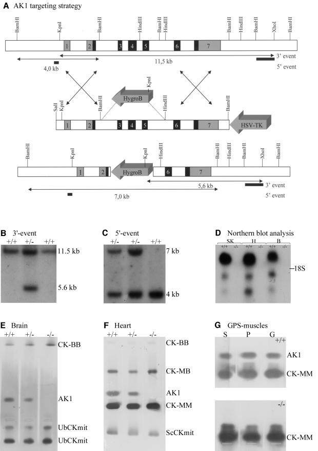
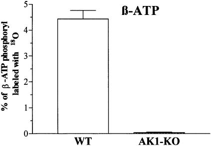
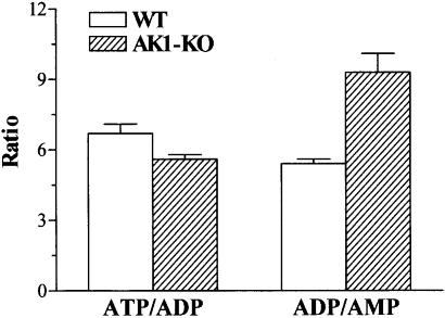
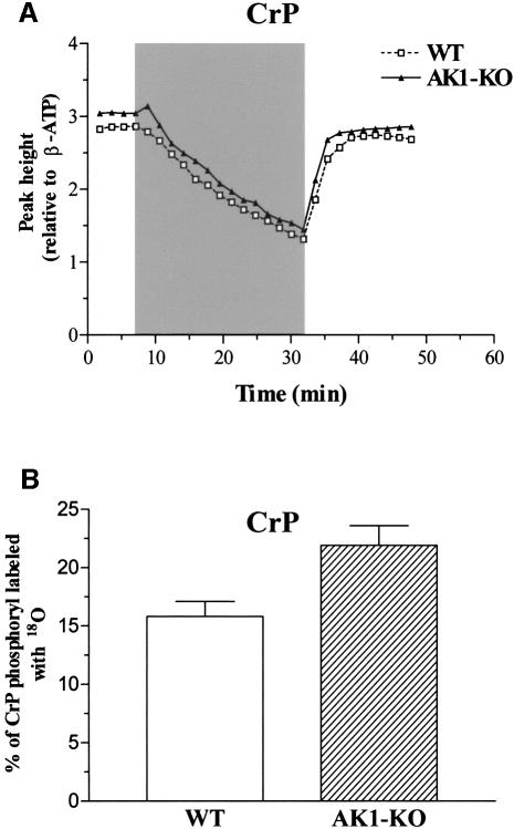
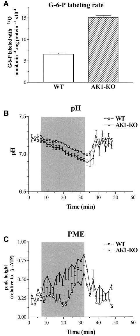
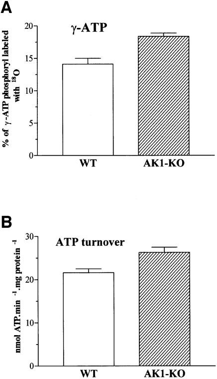
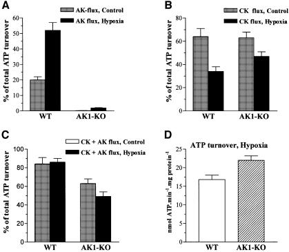
Efficient cellular energy homeostasis is a critical determinant of muscle performance, providing evolutionary advantages responsible for species survival. Phosphotransfer reactions, which couple ATP production and utilization, are thought to play a central role in this process. Here, we provide evidence that genetic disruption of AK1-catalyzed ss-phosphoryl transfer in mice decreases the potential of myofibers to sustain nucleotide ratios despite up-regulation of high-energy phosphoryl flux through glycolytic, guanylate and creatine kinase phosphotransfer pathways. A maintained contractile performance of AK1-deficient muscles was associated with higher ATP turnover rate and larger amounts of ATP consumed per contraction. Metabolic stress further aggravated the energetic cost in AK1(-/-) muscles. Thus, AK1-catalyzed phosphotransfer is essential in the maintenance of cellular energetic economy, enabling skeletal muscle to perform at the lowest metabolic cost.


Figures

None
Fig. 1. (A) Targeted mutagenesis of AK1 with wild-type (top) and mutant (bottom) AK1 genes shown after homologous recombination. Exons: shaded or black; introns or extragenic regions: white. In the targeting vector (middle), a selectable HygroB cassette replaces the 5.5 kb BamHI–HindIII gene fragment (exons 3–5) that encodes the ATP-binding domain of AK1. A HSV-tk cassette was fused to the right arm of homology to facilitate selection of properly targeted ES clones. Lengths of restriction fragments (arrowed lines) from wild-type and mutant alleles are indicated. (B and C) Southern blot of genomic DNAs from wild-type and mutated E14 ES clones. DNAs were cleaved with KpnI–XhoI or BamHI restriction enzymes, resolved by agar gel electrophoresis, and blot-analyzed by hybridization with probes (indicated in A) that discriminate between recombination events at the 3′- (B) and 5′- (C) segments of homology. Diagnostic KpnI–XhoI digestion yields 11.5 and 5.6 kb fragments, whereas BamHI digestion results in 4 and 7 kb fragments for wild-type and mutant alleles. (D) Northern blot analysis of RNAs from wild-type and AK1–/– tissues. Blots with equivalent amounts of total RNA (10 µg) from skeletal muscle (SK), heart (H) and brain (B) probed with AK1 cDNA. Note the absence of AK1 mRNA in homozygous mutants. (E, F and G) Zymogram analysis of homogenates from brain (B), heart (H) and gastrocnemius (G)–plantaris (P)–soleus (S) muscle of wild-type, AK1–/– (homozygous) and AK1+/– (heterozygous) mice. AK1 and CK isoenzymes were separated by agarose gel electrophoresis under native conditions and their migration positions revealed by activity staining. Note the complete absence of staining at the AK1 position for AK1-deficient mice.
None
Fig. 2. Absence of AK phosphotransfer in AK1-knockout skeletal muscle. β-ATP phosphoryl oxygens replaced with 18O, as an indicator of AK-catalyzed phosphotransfer, in wild-type (WT; n = 6) and AK1-knockout (AK1-KO; n = 6) GPS muscle.
None
Fig. 3. Adenine nucleotide ratios in AK1-knockout skeletal muscle. ATP/ADP and ADP/AMP ratios measured in wild-type (n = 6) and AK1-knockout (n = 6) GPS muscle paced for 3 min at 2 Hz.
None
Fig. 4. Creatine phosphate levels and CK phosphotransfer in AK1-knockout skeletal muscle. (A) Levels of CrP in wild-type and AK1–/– mice, analyzed by 31P-NMR and expressed as resonance peak heights normalized to the mean value of the β-ATP intensity of the first four spectra. Series were recorded every 108 s (90° pulses) before, during and after ischemia of the lower hind limb muscles in wild-type (dashed line, open squares) and AK1-deficient (solid line, closed triangles) mice. After four reference spectra, an ischemic period of 25 min (shaded area) was introduced succeeded by a recovery of 16 min. (B) Increased CK-catalyzed CrP turnover in AK1-knockout muscle. Percentage of CrP-phosphoryl oxygens replaced with 18O, as an indicator of CK-catalyzed phosphotransfer, in wild-type (n = 6) and AK1-knockout (n = 6) GPS muscle, determined by mass spectrometry.
None
Fig. 5. (A) Increased hexokinase-catalyzed G-6-P turnover in AK1-knockout muscle. Glucose-6-phosphate phosphoryl oxygens replaced with 18O, as an indicator of glycolytic phosphotransfer, in wild-type (n = 6) and AK1-knockout (n = 6) GPS muscle. (B and C) Intracellular pH (B) and PME levels (C) in wild-type (WT) and AK1–/– mice, analyzed by 31P-NMR and expressed in pH units or as resonance peak heights normalized to the mean value of the β-ATP intensity of the first four spectra. Series were recorded before, during and after ischemia of the lower hind limb muscles in wild-type (dashed line, open squares) and AK1-deficient (solid line, closed triangles) mice. After four reference spectra, an ischemic period of 25 min was introduced succeeded by a recovery of 16 min. Note a faster pH decrease and increased PME levels during the ischemic period in AK1–/– compared with wild-type mice.
None
Fig. 6. Guanine nucleotide metabolism in AK1-knockout skeletal muscle. (A) The GTP/GDP and ATP/GTP ratios were measured in wild-type (n = 6) and AK1-knockout (n = 6) GPS muscle paced for 3 min at 2 Hz. (B) Increased γ-GTP phosphate turnover in AK1-knockout skeletal muscle. Percentage of γ-GTP phosphoryl oxygens replaced with 18O, as an indicator of enzyme activity catalyzing GTP production, in wild-type (n = 6) and AK1-knockout (n = 6) GPS muscle. (C) Increased guanylate kinase-catalyzed phosphotransfer in AK1-knockout skeletal muscle. Percentage of β-GTP/GDP phosphoryl oxygens replaced with 18O, as an indicator of guanylate kinase phosphotransfer, in wild-type (n = 6) and AK1-knockout (n = 6) GPS muscle.
None
Fig. 7. Increased ATP turnover in AK1-knockout skeletal muscle. (A) Increased γ-ATP phosphate turnover in AK1-knockout GPS. Percentage of γ-ATP phosphoryl oxygens replaced with 18O, as an indicator of ATP synthesis rate, in wild-type (n = 6) and AK1-knockout (n = 6) GPS muscle. (B) Increased ATP turnover in AK1-knockout skeletal muscle. Total ATP turnover, obtained from 18O incorporation into major high-energy phosphoryls, in wild-type (n = 6) and AK1-knockout (n = 6) GPS muscle.
None
Fig. 8. Aberrant redistribution of phosphotransfer flux in AK1-knockout skeletal muscle in response to hypoxia. (A) Hypoxia markedly elevates AK-catalyzed flux in wild-type, but not in AK1-knockout GPS. AK-catalyzed phosphotransfer, expressed as a percentage of total cellular ATP turnover, in wild-type (n = 6) and AK1-knockout (n = 6) GPS muscle. (B) Increased contribution of CK-catalyzed phosphotransfer in hypoxic AK1-deficient compared with hypoxic wild-type GPS. Creatine kinase-catalyzed phosphotransfer, expressed as a percentage of total ATP turnover, in wild-type (n = 6) and AK1-knockout (n = 6) GPS muscle. (C) Contribution of combined AK- and CK-catalyzed phosphotransfer to cellular ATP turnover is reduced in normoxic and hypoxic AK1-knockout GPS. The sum of AK- and CK-catalyzed phosphotransfer is expressed as a percentage of total ATP turnover measured by the [18O]phosphoryl labeling technique. (D) ATP turnover rates in wild-type and AK1-knockout GPS under hypoxia. Hypoxia was induced by KCN (2 mM), an inhibitor of mitochondrial respiration.

References

    1. Bandlow W., Strobel,G., Zoglowek,C., Oechsner,U. and Magdolen,V. (1988) Yeast adenylate kinase is active simultaneously in mitochondria and cytoplasm and is required for non-fermentative growth. Eur. J. Biochem., 178, 451–457. - PubMed
    1. Bessman S.P. and Carpenter,C.L. (1985) The creatine–creatine phosphate energy shuttle. Annu. Rev. Biochem., 54, 831–862. - PubMed
    1. Collavin L., Lazarevic,D., Utrera,R., Marzinotto,S., Monte,M. and Schneider,C. (1999) wt p53 dependent expression of a membrane-associated isoform of adenylate kinase. Oncogene, 18, 5879–5888. - PubMed
    1. de Haan A., Jones,D.A. and Sargeant,A.J. (1989) Changes in velocity of shortening, power output and relaxation rate during fatigue of rat medial gastrocnemius muscle. Pflugers Arch., 413, 422–428. - PubMed
    1. Dzeja P.P. and Terzic,A. (1998) Phosphotransfer reactions in the regulation of ATP-sensitive K+ channels. FASEB J., 12, 523–529. - PubMed

Publication types

MeSH terms

LinkOut - more resources