. 2000 Dec 1;19(23):6382-91.
doi: 10.1093/emboj/19.23.6382.

The peroxisomal membrane protein Pex13p shows a novel mode of SH3 interaction

Affiliations

The peroxisomal membrane protein Pex13p shows a novel mode of SH3 interaction

P Barnett et al. EMBO J. .

Abstract

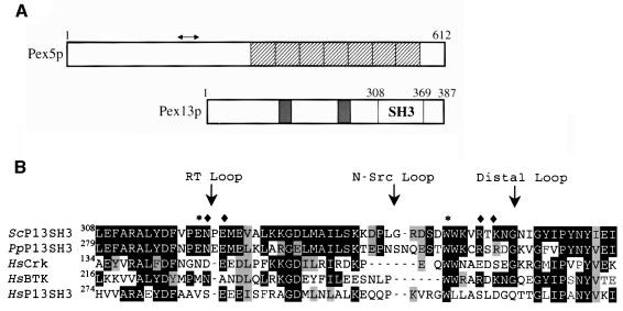
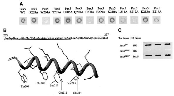
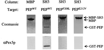
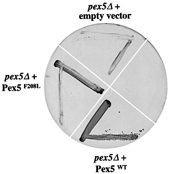
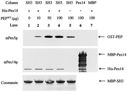
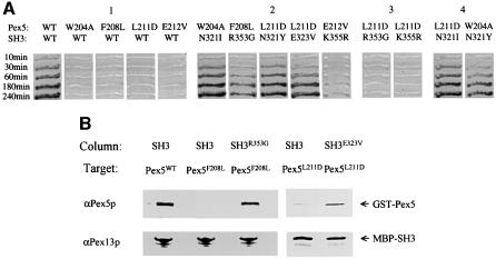
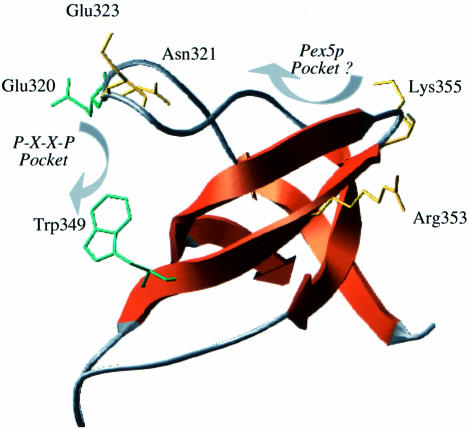
Src homology 3 (SH3) domains are small non-catalytic protein modules capable of mediating protein-protein interactions by binding to proline-X-X-proline (P-X-X-P) motifs. Here we demonstrate that the SH3 domain of the integral peroxisomal membrane protein Pex13p is able to bind two proteins, one of which, Pex5p, represents a novel non-P-X-X-P ligand. Using alanine scanning, two-hybrid and in vitro interaction analysis, we show that an alpha-helical element in Pex5p is necessary and sufficient for SH3 interaction. Sup pressor analysis using Pex5p mutants located in this alpha-helical element allowed the identification of a unique site of interaction for Pex5p on the Pex13p-SH3 domain that is distinct from the classical P-X-X-P binding pocket. On the basis of a structural model of the Pex13p-SH3 domain we show that this interaction probably takes place between the RT- and distal loops. Thus, the Pex13p-SH3-Pex5p interaction establishes a novel mode of SH3 interaction.


Figures

None
Fig. 1. (A) Schematic representation of the domain structure of Pex5p and Pex13p. Shown for Pex5p are the seven TPR repeats (hatched boxes) and the region involved in Pex13p-SH3 binding (arrow). For Pex13p, the two predicted transmembrane regions (filled boxes) and the SH3 domain are indicated. (B) Alignment of the SH3 domains from: Saccharomyces cerevisiae Pex13p (ScP13SH3) P80667, Pichia pastoris Pex13p (PpP13SH3) Q92266, human Crk (HsCrk) P46108, human BTK (HsBTK) Q06187 and human Pex13p (HsP13SH3) Q92968. Sequences were aligned using ClustalX and manual fitting. White text on a black background denotes a sequence residue identity and black text on a grey background a similarity. Positions of the RT-loop, N-Src and Distal loop are indicated with an arrow. The RT-loop residue Glu320 and the conserved Trp349, both important in P-X-X-P ligand recognition, are marked with an asterisk. Residues that were found mutated in the suppressor screen (see Figure 6) are marked with a diamond.
None
Fig. 2. Analysis of the Pex5p–Pex13p-SH3 interaction. (A) Two-hybrid analysis of Pex5p alanine scan mutants. Wild type or Pex5p mutants fused to the TA domain were co-transformed with DB Pex13p-SH3 to PCY2, and assayed for β-galactosidase activity using a filter assay. Black indicates a strong interaction, white shows no interaction and grey indicates a weakened interaction. (B) Secondary structural model of the Pex13p-SH3 binding element from Pex5p. The model was generated in Swiss-PDB viewer and side chains are depicted in default torsion angles. The sequence at the top of the figure shows the region of Pex5p used for in vitro binding studies (Figure 3). Amino acids tested in the alanine scan appear in italic. The underlined sequence is represented in the helical model. The side chains of residues affecting the interaction of the Pex5p with Pex13p-SH3 are marked on the helix and labelled. (C) Two-hybrid analysis of Pex5p Lys210Pro mutant. Wild-type Pex5p or mutant Pex5p Lys210Pro fused to the TA domain were co-transformed with DB Pex13p-SH3 into PCY2, and assayed for β-galactosidase activity using a filter assay. As a control, TA Pex5p Lys210Pro was also tested against DB Pex14p. Shown are three independent yeast transformants.
None
Fig. 3. In vitro binding experiments of Pex5p peptides and Pex13p-SH3. GST-fused Pex5p peptide (PEPWT) (residues 203–227) or GST-fused Pex5p peptides possessing either the Phe208Leu mutation (PEPF208L) or the Glu212Val mutation (PEPE212V) (100 µg each) were passed over affinity columns loaded with 250 µl of cleared lysate containing either MBP alone or MBP-fused Pex13p-SH3 (SH3). After appropriate washing, proteins were eluted from the column with maltose. Eluates were subjected to SDS–PAGE and gels were stained with Coomassie (top panel) or blotted and probed with antibodies against Pex5p (lower panel). Protein bands are appropriately labelled on the right-hand side of the figure.
None
Fig. 4. In vivo analysis of Pex5p Phe208Leu. pex5Δ cells were transformed with wild-type Pex5p (Pex5WT), Pex5p Phe208Leu (pex5F208L) or with an empty plasmid. Cells were grown to mid-log phase in liquid medium containing 0.3% glucose and plated on oleate medium. Plates were incubated at 28°C and photographed after 7 days.
None
Fig. 5. In vitro competition assay. Lanes 1–5, constant amounts of E.coli lysates containing MBP-SH3 (SH3) and His6-Pex14p (HisPex14p) (100 µl cleared lysate of each) were mixed with increasing amounts of purified GST–Pex5p peptide fusion (PEPWT) (0–100 µg fusion peptide). In lane 5, His6-Pex14p was omitted from the incubation. Lane 6, 100 µl of E.coli lysate containing MBP-Pex14p (Pex14) were mixed with 100 µg of Pex5p peptide fusion. Lane 7, 100 µl of lysate containing MBP were mixed with His6-Pex14p (100 µl) and Pex5p peptide fusion (100 µg). After incubation the mixtures were loaded onto an amylose column then washed and eluted. Eluates were analysed by SDS–PAGE and stained with Coomassie Blue (bottom panel) or blotted and probed with antibodies (upper panels) specific for Pex5p and Pex14p.
None
Fig. 6. Analysis of Pex13p-SH3 suppressor mutants. (A) Two-hybrid analysis. PCY2 was co-transformed with plasmids encoding the proteins as indicated and tested for β-galactosidase activity using a filter assay. Filters were imaged at specific time intervals to convey relative strengths of interaction. Panel 1 shows the interaction of wild-type Pex5p and various Pex5p mutants with Pex13p-SH3 wild type. Panel 2 shows the interaction between Pex5p mutants and their corresponding Pex13p-SH3 suppressors. Panel 3 displays an example of the allele specificity of the suppressors. Panel 4 shows the dual nature of the suppressors picked up for Pex5p Trp204Ala and Pex5p Leu211Asp at the same position on the SH3 domain. Note that Pex5p Leu211Asp apparently has no preference for Ile or Tyr at position 321 of the SH3 domain, whereas Pex5p Trp204Ala displays a preference for an Ile at this position. (BIn vitro analysis. Wild-type Pex5p (Pex5WT) or mutant Pex5p (Pex5F208L) fused to GST was passed over affinity columns loaded with either MBP-SH3 (SH3) or MBP-SH3 Arg353Gly (SH3R353G). Similarly, Pex5p Leu211Asp (Pex5L211D) fused to GST was passed over affinity columns loaded with either MBP-SH3 (SH3) or MBP-SH3 Glu323Val (SH3E323V). Washing, elution and analysis of the eluates were carried out as described in the legend to Figure 3. Eluates were analysed by SDS–PAGE and western blotting using antibodies specific for Pex5p and Pex13p-SH3.
None
Fig. 7. Structural model of the Pex13p-SH3 domain. Structural model showing the secondary structural elements of the Pex13p-SH3 domain. Side chains in green specifically affect association of Pex14p. Side chains in yellow are residues that were picked up in the suppressor screen. These residues do not directly affect Pex14p association. The position of the P-X-X-P binding pocket important for Pex14p association, and the possible Pex5p binding cleft are marked.

Similar articles

Cited by

References

    1. Albertini M., Rehling,P., Erdmann,R., Girzalsky,W., Kiel,J.A., Veenhuis,M. and Kunau,W.H. (1997) Pex14p, a peroxisomal membrane protein binding both receptors of the two PTS-dependent import pathways. Cell, 89, 83–92. - PubMed
    1. Arold S., O’Brien,R., Franken,P., Strub,M.P., Hoh,F., Dumas,C. and Ladbury,J.E. (1998) RT loop flexibility enhances the specificity of Src family SH3 domains for HIV-1 Nef. Biochemistry, 37, 14683–14691. - PubMed
    1. Blatch G.L. and Lassle,M. (1999) The tetratricopeptide repeat: a structural motif mediating protein–protein interactions. BioEssays, 21, 932–939. - PubMed
    1. Bottger G., Barnett,P., Klein,A.T.J., Kragt,A., Tabak,H.F. and Distel,B. (2000) Saccharomyces cerevisiae PTS1 receptor Pex5p interacts with the SH3 domain of the peroxisomal membrane protein Pex13p in an unconventional, non-PXXP-related manner. Mol. Biol. Cell., in press. - PMC - PubMed
    1. Brocard C., Kragler,F., Simon,M.M., Schuster,T. and Hartig,A. (1994) The tetratricopeptide repeat-domain of the PAS10 protein of Saccharomyces cerevisiae is essential for binding the peroxisomal targeting signal-SKL. Biochem. Biophys. Res. Commun., 204, 1016–1022. - PubMed

Publication types

MeSH terms