. 2000 Dec 1;19(23):6408-18.
doi: 10.1093/emboj/19.23.6408.

Components and dynamics of fiber formation define a ubiquitous biogenesis pathway for bacterial pili

Affiliations

Components and dynamics of fiber formation define a ubiquitous biogenesis pathway for bacterial pili

M Wolfgang et al. EMBO J. .

Abstract

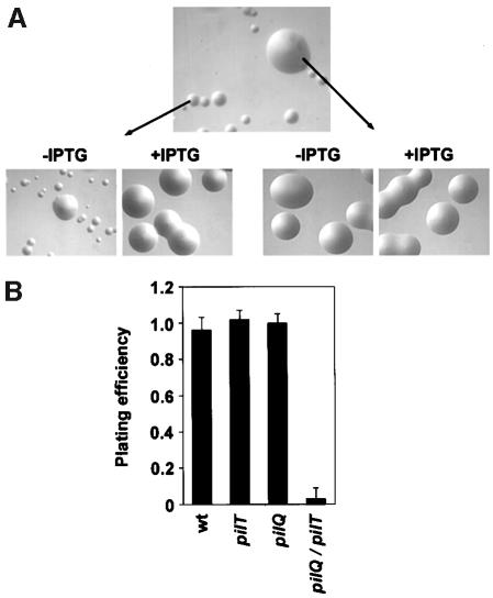
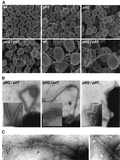
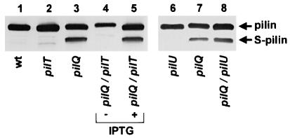
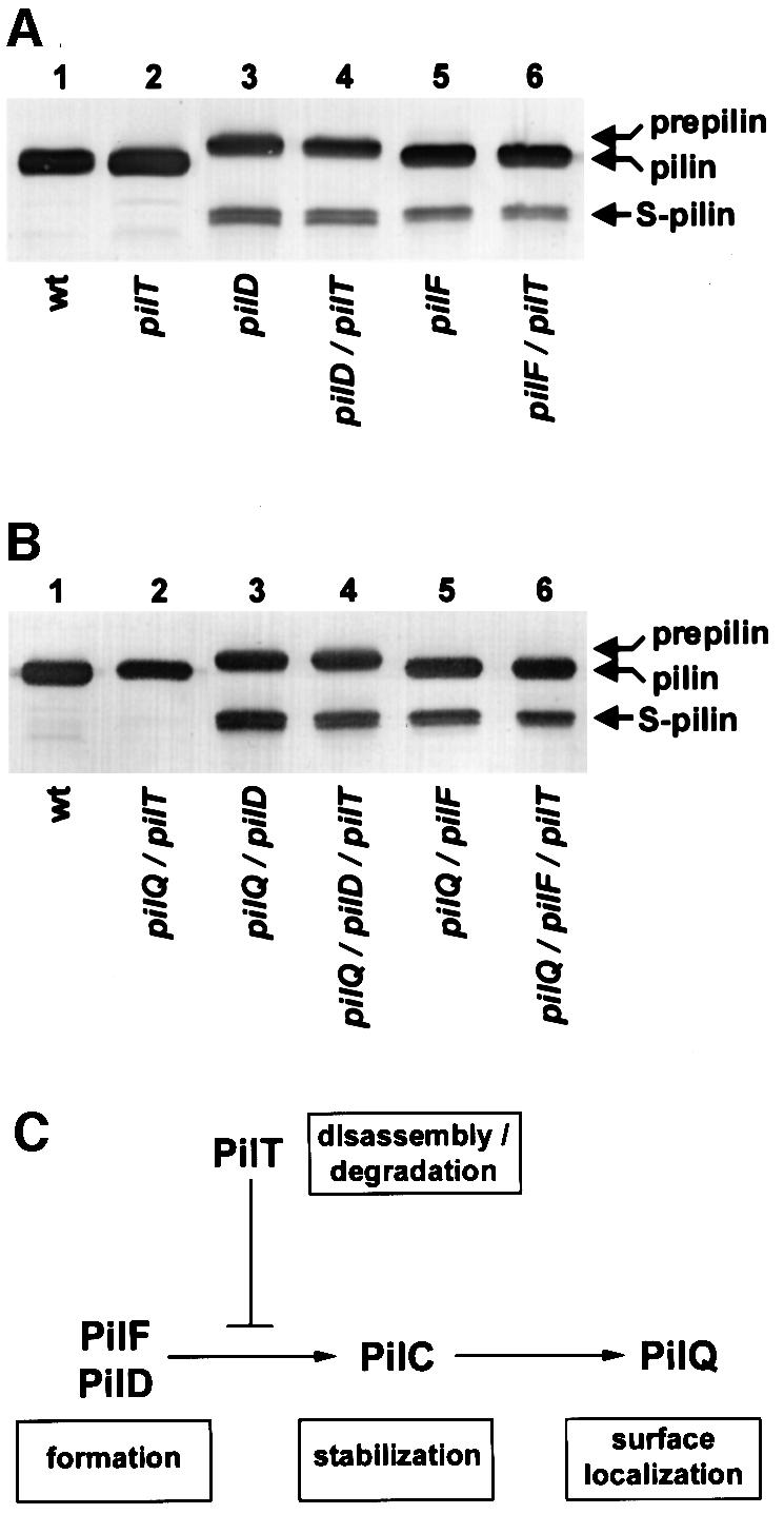
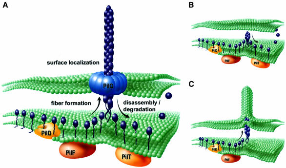
Type IV pili (Tfp) are a unique class of multifunctional surface organelles in Gram-negative bacteria, which play important roles in prokaryotic cell biology. Although components of the Tfp biogenesis machinery have been characterized, it is not clear how they function or interact. Using Neisseria gonorrhoeae as a model system, we report here that organelle biogenesis can be resolved into two discrete steps: fiber formation and translocation of the fiber to the cell surface. This conclusion is based on the capturing of an intermediate state in which the organelle is retained within the cell owing to the simultaneous absence of the secretin family member and biogenesis component PilQ and the twitching motility/pilus retraction protein PilT. This finding is the first demonstration of a specific translocation defect associated with loss of secretin function, and additionally confirms the role of PilT as a conditional antagonist of stable pilus fiber formation. These findings have important implications for Tfp structure and function and are pertinent to other membrane translocation systems that utilize a highly related set of components.


Figures

None
Fig. 1. Simultaneous loss of PilQ and PilT expression leads to defects in growth reflected in altered colony size and plating efficiencies. (A) Neisseria gonorrhoeae colonies photographed after 24 h growth on solid agar at a magnification of 30× using a stereomicroscope. Top panel: strain MW11 (pilQ::mTncm21, pilTind). Lower left panel: relief of the growth defect in MW11 by derepression of pilT expression (+IPTG). Lower right panel: variants that suppress the growth defect can be isolated based on their ability to yield colonies of normal size, regardless of pilT expression. (B) Effects of pilT derepression on the plating efficiencies of wild-type and mutant N.gonorrhoeae strains. The ratio of colony forming seen after 24 h in the absence and presence of pilT de-repression was determined. Data represent the average of three experiments. Strain designation and genotype for the mutants presented are as follows: wt (N400), pilT (MW4, pilTind), pilQ (GQ21, pilQ::mTncm21) and pilQ/pilT (MW11, pilQ::mTncm21, pilTind).
None
Fig. 2. Electron microscopic analysis of pilQ/pilT mutants shows altered cell surface structures comprised of membrane-bound pilus filaments. (A) Scanning electron micrographs of wild-type and mutant N.gonorrhoeae strains. wt (N400); pilT (MW4, pilTind); pilQ (GQ21, pilQ::mTncm21); pilQ/pilT (MW11, pilQ::mTncm21, pilTind). In the lower center and lower right panels, wt and pilQ/pilT micrographs are at 50 000× magnification; all others are at 25 000×. Individual N.gonorrhoeae cells are ∼1 µm in diameter. (B) Transmission electron micrographs showing membrane-bound pilus fibers in a pilQ/T mutant (strain MW11). Note that bulges seen in the membranous protrusions contain coiled fibers detected by TEM (center and right panels) and that they correspond to analogous structures seen in SEM. Micrographs are taken at a magnification of 90 000× and inset panels show digitally enlarged images at a 3× higher magnification. (C) Immunolabeling of fibers associated with disrupted blebs with antiserum raised against purified pili (135 000×).
None
Fig. 3. PilT is required for fiber subunit degradation in pilQ mutants. Abolition of fiber expression and associated growth defects in the pilQ/pilT mutant by de-repression of pilT expression leads to subunit degradation. An immunoblot of whole-cell lysates probed with the pilin-specific monoclonal antibodies (mAb MC02) is shown. Lane 1, wild-type (N400, recA6); lane 2, pilT (MW4, pilTind); lane 3, pilQ (GQ21, pilQ::mTncm21); lanes 4 and 5, pilQ/pilT (MW11, pilQ::mTncm21, pilTind); lane 6, pilU (MW35, pilU::kan); lane 7, pilQ (GQ21, pilQ::mTncm21); lane 8, pilQ/pilU (MW36, pilQ::mTncm21, pilU::kan). (+) denotes lysates derived from strains propagated in the presence of pilT expression (plus IPTG). S-pilin denotes the migration of the truncated species of PilE lacking the first 39 residues present in the mature molecule.
None
Fig. 4. Variants that suppress the growth defect associated with the lack of PilQ and PilT show alterations in the expression, degradation and structure of the pilin subunit. (A) Immunoblot analysis of pilin subunits expressed by variants derived from the pilQ/pilT mutant (strain MW11, pilQ::mTncm21, pilTind) that no longer show a growth defect. Pilin-specific mAb MC02 was used to probe whole-cell lysates. Lane 1, wild-type (N400, recA6); lane 2, pilQ/pilT (MW11, pilQ::mTncm21, pilTind); lanes 3–14, variants isolated from MW11 (strains MW12–MW23, respectively). (B) Variants that suppress the pilQ/pilT-associated growth defect show multiple changes in the primary structure of pilin. The predicted amino acid sequence of pilin, expressed from pilE (designated pilEwt) of the parental pilQ/pilT strain is shown. Changes in the predicted primary structure of variant pilins, based on DNA sequence, are indicated. Periods (⋅) denote identical residues and dashes (–) represent gaps in the sequence. N-terminal sequences are not shown, as no changes were detected. No sequence is presented for MW19 as pilE was deleted in this strain.
None
Fig. 5. Variant pilE alleles that arise in association with suppression of the pilQ/pilT growth defect are intrinsically defective in Tfp biogenesis. (A) Schematic diagram outlining the approach used to analyze variant pilE alleles. Strain MW24 was constructed such that expression from endogenous pilE was placed under the control of a regulated promoter. The chromosomal organization for this strain is shown at the top. The expression of altered pilE alleles arising in the pilQ/pilT background was analyzed following cloning and recombination into an ectopic site within the gonococcal iga locus of strain MW24, as depicted at the bottom. In the absence of induction, pilin is produced solely from the ectopically expressed allele. (B) Upper panel: immunoblot analysis of pilin, expressed from the altered pilE alleles, using the pilin-specific mAb MC02. Lower panel: Coomassie Blue-stained SDS–polyacrylamide gel showing the relative amounts of pilin subunit in purified pilus preparations. Lane 1, wild-type (N400, recA6); lane 2, pilE (MW24, pilEind); lanes 3–12, designated pilE alleles expressed ectopically in the pilEind background (strains MW25–MW34, respectively).
None
Fig. 6. PilT does not influence PilE degradation in pilD and pilF mutants or double mutants simultaneously lacking PilQ. PilE degradation was assessed by immunoblotting of whole-cell lysates using the pilin-specific mAb MC02. Prepilin denotes the migration of the unprocessed PilE, while S-pilin denotes the migration of the truncated species of PilE lacking the first 39 residues present in the mature molecule. (A) PilT does not alter PilE stability in pilD and pilF mutants. Lane 1, wild-type (N400, recA6); lane 2, pilT (MW4, pilTind); lane 3, pilD (GDClaI–XhoI, pilDfs); lane 4, pilD/pilT (MW37, pilDfs, pilTind); lane 5, pilF (GF2, pilF::mTnerm2); lane 6, pilF/pilT (MW38, pilF::mTnerm2, pilTind). (B) Epistastic relationships of pilD, pilF and pilQ with regard to PilT-dependent PilE degradation. Lane 1, wild-type (N400, recA6); lane 2, pilQ/pilT (MW39, pilQind, pilTind); lane 3, pilQ/pilD (MW40, pilQind, pilDfs); lane 4, pilQ/pilD/pilT (MW41, pilQind, pilDfs, pilTind); lane 5, pilQ/pilF (MW42, pilQind, pilF::mTnerm2); lane 6, pilQ/pilF/pilT (MW43, pilQind, pilF::mTnerm2, pilTind). (C) Inferred order of action of components in the Tfp biogenesis pathway. Shown is a diagrammatic pathway summarizing the activities of Tfp biogenesis components in fiber formation, stabilization and surface localization as determined from this study and prior results (Wolfgang et al., 1998b). Note that although PilT is shown to impact antagonistically on the pathway downstream of PilD and PilF, this does not relate directly to its actual physical localization in the cell or the site where it may function.
None
Fig. 7. Components and dynamics of Tfp biogenesis. (A) Modeling of the conditions operating in wild-type cells in which a growing Tfp fiber is translocated to the cell surface via the secretin PilQ. This configuration provides a mechanism by which a common machinery at the inner membrane–periplasm interface (inferred as fiber formation mediated by PilF and antagonism of stable fiber formation/depolymerization mediated by PilT) can lead to pilus retraction by virtue of acting on a contiguous pilus filament. (B) Model depicting the events in mutants lacking the secretin protein PilQ. Following processing of pilin PilE (purple) by PilD and the function served by the GspE/PulE family member PilF, stable fiber formation is antagonized by the action of the related family member PilT. Due to physical restriction of fiber translocation to the cell surface, the equilibrium between fiber growth and retraction/depolymerization is shifted such that no fibers are seen. (C) As in (B) but in the absence of PilT, stable fiber formation leads to membranous extrusions generated by protrusive force and defects in cell viability. Suppression of formation: the membranous extrusions and growth defects can occur by loss of PilD or PilF function as well as mutations in pilE, encoding the fiber subunit.

Similar articles

Cited by

References

    1. Abraham V.C., Krishnamurthi,V., Taylor,D.L. and Lanni,F. (1999) The actin-based nanomachine at the leading edge of migrating cells. Biophys. J., 77, 1721–1732. - PMC - PubMed
    1. Bitter W., Koster,M., Latijnhouwers,M., de Cock,H. and Tommassen,J. (1998) Formation of oligomeric rings by XcpQ and PilQ, which are involved in protein transport across the outer membrane of Pseudomonas aeruginosa. Mol. Microbiol., 27, 209–219. - PubMed
    1. Bradley D.E. (1980) A function of Pseudomonas aeruginosa PAO polar pili: twitching motility. Can. J. Microbiol., 26, 146–154. - PubMed
    1. Crago A.M. and Koronakis,V. (1998) Salmonella InvG forms a ring-like multimer that requires the InvH lipoprotein for outer membrane localization. Mol. Microbiol., 30, 47–56. - PubMed
    1. Dougherty B.A. and Smith,H.O. (1999) Identification of Haemophilus influenzae Rd transformation genes using cassette mutagenesis. Microbiology, 145, 401–409. - PubMed

Publication types

MeSH terms