. 2000 Dec 1;19(23):6419-26.
doi: 10.1093/emboj/19.23.6419.

SRP-dependent co-translational targeting and SecA-dependent translocation analyzed as individual steps in the export of a bacterial protein

Affiliations

SRP-dependent co-translational targeting and SecA-dependent translocation analyzed as individual steps in the export of a bacterial protein

C Neumann-Haefelin et al. EMBO J. .

Abstract

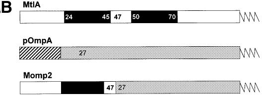
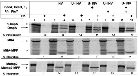
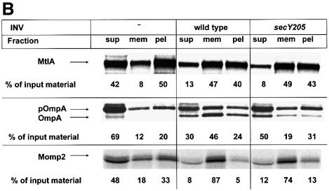
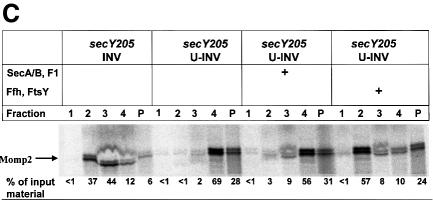
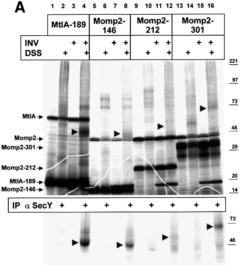
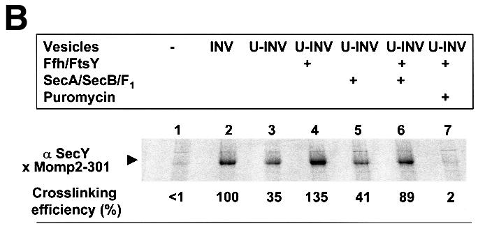
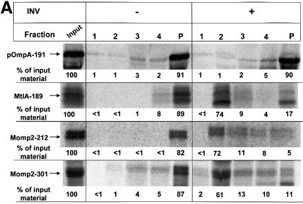
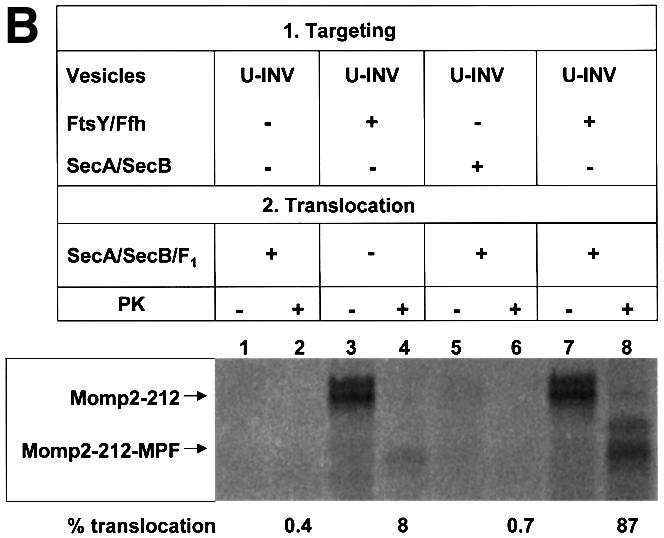
Recently it has been recognized that the signal recognition particle (SRP) of Escherichia coli represents a specific targeting device for hydrophobic inner membrane proteins. It has remained unclear, however, whether the bacterial SRP functions in concert with SecA, which is required for the translocation of secretory proteins across the inner membrane. Here, we have analyzed a hybrid protein constructed by fusing the signal anchor sequence of an SRP-dependent inner membrane protein (MtlA) to the mature part of an exclusively SecA-requiring secretory protein (OmpA). We show that the signal anchor sequence of MtlA confers the novel properties onto nascent chains of OmpA of being co-translationally recognized and targeted to SecY by SRP. Once targeted to SecY, ribosome-associated nascent chains of the hybrid protein, however, remain untranslocated unless SecA is present. These results indicate that SRP and SecA cooperate in a sequential, non-overlapping manner in the topogenesis of those membrane proteins which, in addition to a signal anchor sequence, harbor a substantial hydrophilic domain to be translocated into the periplasm.


Figures

None
Fig. 1. Nascent chains of Momp2, a hybrid between the polytopic membrane protein MtlA and the secretory protein pOmpA, bind to both Ffh and trigger factor. (A) pOmpA, MtlA and Momp2 were synthesized in vitro by coupled transcription–translation of plasmids pDMB, p717MtlA-B and pMomp2, respectively. The addition of appropriate antisense oligodeoxynucleotides gave rise to a 125 amino acid N-terminal fragment of pOmpA (pOmpA-125), a 189 amino acid fragment of MtlA (MtlA-189) and a 146 amino acid fragment of Momp2 (Momp2-146). Co-synthesis of some full-length products (MtlA, OmpA and Momp2) was observed under these conditions. [35S]methionine-labeled translation products were precipitated with trichloroacetic acid (TCA), separated by SDS–PAGE (7–17% acrylamide) and visualized by phosphoimaging. Where indicated, Ffh (2 ng/µl) was present during synthesis. Cross-linking with DSS was followed by centrifugation through a sucrose cushion in a TLA-100.2 Beckmann rotor at 70 000 r.p.m. for 60 min at 4°C (Behrmann et al., 1998; Beck et al., 2000) and immunprecipitation (IP) using anti-Ffh and anti-trigger factor (TIG) antibodies. Molecular masses are indicated to the right. Cross-linking products with trigger factor (*) and Ffh (x) are indicated. (B) Construction of the hybrid protein Momp2 from MtlA and OmpA. For experimental details, see Materials and methods. Black boxes represent predicted transmembrane segments of MtlA (Sugiyama et al., 1991), with the preceeding and following amino acids each indicated in white. Numbering starts with the first N-terminal amino acid, with that of MtlA sequences given in bold. Only an arbitrarily long N-terminal part of each protein is drawn. The signal sequence of pOmpA is depicted as a hatched area, and the mature sequence as a gray box.
None
Fig. 2. Translocation of Momp2 into membrane vesicles is achieved solely by SecA, SecB and the F1-ATPase. pOmpA, MtlA and Momp2 were synthesized in vitro in the presence of the components indicated at the top (INV, E.coli inside-out inner membrane vesicles; U-INV, urea-extracted INV). SecA (80 ng/µl), SecB (40 ng/µl), F1-ATPase (40 ng/µl), Ffh (2 ng/µl) and FtsY (20 ng/µl) were added as specified. Translation products were either precipitated directly with TCA or only after incubation with 0.5 mg/ml proteinase K (PK) for 20 min at 25°C. Indicated are the positions of the precursor (pOmpA) and the signal sequence-free form of OmpA, of full-length MtlA and its fragment resistant towards proteinase K (MtlA-MPF), and of Momp2 and its fragment resistant towards proteinase K (Momp2-MPF).
None
Fig. 3. Translocation of Momp2 requires a functional SecY–SecA interaction but not its membrane association. (A) pOmpA, MtlA and Momp2 were synthesized in vitro in the presence of INV buffer, wild-type INVs or secY205 INVs and the endogenous amounts of SecA, SecB, F1-ATPase, Ffh and FtsY. The percentage of translocation and integration was calculated by quantitation of the radioactivity measured in individual protein bands using phosphoimaging. The indicated percentage of OmpA translocation equals the ratio between radioactivity in the bands of pOmpA and OmpA after and before proteolytic digestion. The percentage of MtlA integration was calculated by the ratio between MtlA-MPF and MtlA, corrected for the loss of [35S]methionine residues during cleavage by proteinase K. The translocation of Momp2 equals the ratio between Momp2-MPF and Momp2. (B) As in (A), but membrane association was analyzed by subfractionation of the translation products on a two-step sucrose gradient. Radioactivity of the three subfractions was quantified and the sum set at 100%. (C) Flotation analyses of full-length Momp2 in the presence of secY205 INVs or secY205 U-INVs. In vitro synthesis of Momp2 was performed in the presence of the indicated components (see Figure 2 for the concentrations used) and the reaction mixture was subsequently separated by a flotation gradient centrifugation. Following centrifugation, 4 × 100 µl fractions were withdrawn from the top of the gradient and, after precipitation with TCA, separated by 15% SDS–PAGE. The pellet fraction was dissolved directly in loading buffer. Fractions 2 and 3 correspond to the membrane fraction.
None
Fig. 4. Co-translational targeting of Momp2 RNCs to SecY, which is mediated by SRP. (A) Nascent chains of MtlA and Momp2 of the indicated lengths were synthesized in vitro (cf. Figure 1) in the presence of the endogenous amounts of SecA, SecB, F1-ATPase, Ffh and FtsY. When indicated, inside-out inner membrane vesicles (INVs) were present during synthesis and samples were treated post-translationally with DSS. Immunoprecipitation of the samples in the even-numbered lanes using anti-SecY polyclonal antibodies is shown below. (B) RNCs of Momp2-301 were synthesized in vitro in the presence of the indicated components. Nascent chains in lane 7 were treated with 0.8 mM puromycin for 10 min at 37°C before immunoprecipitation.
None
Fig. 5. SRP-dependent targeting and SecA-dependent translocation are distinct steps in the translocation of Momp2 nascent chains. (A) Flotation analyses of ribosome-associated nascent chains (RNCs) of OmpA, MtlA and Momp2 in the absence or presence of membrane vesicles (INV). After in vitro synthesis in the presence of SecA, SecB, Ffh and FtsY, RNCs were incubated with INV buffer or INVs for 15 min at 37°C. For experimental details, see Materials and methods and Figure 3. (B) Momp2 RNCs were synthesized in the presence of urea-treated INVs (U-INV). FtsY, Ffh, SecA and SecB were present during synthesis as indicated. After flotation as specified in (A), the INV-containing fraction 2 of each gradient was withdrawn and incubated for 15 min at 37°C in the presence of puromycin to release the ribosome. SecA, SecB and F1-ATPase were present during this incubation. The reaction mixtures were subsequently split in half; one half was precipitated directly with TCA to identify the membrane-associated nascent chains of Momp2, and the other was first digested with proteinase K (PK) before precipitation with TCA to test for complete translocation.
None
Fig. 6. Model of the export of Momp2 as an example of membrane-anchored periplasmic E.coli proteins. (A) Ribosome-associated nascent chains of Momp2 are recognized co-translationally by SRP (Ffh and 4.5S RNA) via their signal anchor sequence. For simplicity, the experimentally verified interaction of nascent Momp2 with trigger factor is not depicted. SecA does not associate with Momp2 at this stage. (B) Following interaction between SRP and its receptor (FtsY), nascent Momp2 is targeted co-translationally to SecY. Again, this step does not require SecA. SecA, therefore, is not an indispensable constituent of the SecY translocon. (C) After the initial insertion into the translocon, SecA subsequently binds to Momp2 in order to promote translocation of the hydrophilic OmpA moiety across the membrane. It is not clear whether the presumed direct contact between ribosome and SecY is loosened at this stage and whether SecA binds first to the polypeptide chain or to SecY. SecG, which is not required for the insertion of inner membrane proteins into the SecY translocon (Koch and Müller, 2000), has been omitted despite its unquestioned involvement in the SecA-dependent translocation step.

References

    1. Beck K., Wu,L.F., Brunner,J. and Muller,M. (2000) Discrimination between SRP- and SecA/SecB-dependent substrates involves selective recognition of nascent chains by SRP and trigger factor. EMBO J., 19, 134–143. - PMC - PubMed
    1. Behrmann M., Koch,H.G., Hengelage,T., Wieseler,B., Hoffschulte,H.K. and Muller,M. (1998) Requirements for the translocation of elongation-arrested, ribosome-associated OmpA across the plasma membrane of Escherichia coli. J. Biol. Chem., 273, 13898–13904. - PubMed
    1. Crowley K.S., Reinhart,G.D. and Johnson,A.E. (1993) The signal sequence moves through a ribosomal tunnel into a noncytoplasmic aqueous environment at the ER membrane early in translocation. Cell, 73, 1101–1115. - PubMed
    1. Crowley K.S., Liao,S., Worrel.,V.E., Reinhart,G.D. and Johnson,A.E. (1994) Secretory proteins move through the endoplasmic reticulum membrane via an aqueous, gated pore. Cell, 78, 461–471. - PubMed
    1. de Gier J.W., Mansournia,P., Valent,Q.A., Phillips,G.J., Luirink,J. and von Heijne,G. (1996) Assembly of a cytoplasmic membrane protein in Escherichia coli is dependent on the signal recognition particle. FEBS Lett., 399, 307–309. - PubMed

Publication types

MeSH terms

Substances

LinkOut - more resources